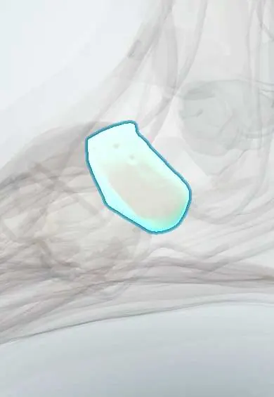
Navicular !
The navicular is a boat-shaped bone on the inner midfoot, at the top of the arch, in front of the talus.
Official name: Navicular bone
Opens in the Durgical app, no sign-up needed.
What it is
A boat-shaped bone on the inner side of the midfoot, between the talus and the three cuneiforms.
What it does
Acts as the keystone of the inner arch. The tibialis posterior tendon attaches to it.
Key facts
- Joints with
- Talus (head); three cuneiforms; sometimes cuboid
- Landmarks
- Navicular tuberosity (main insertion of tibialis posterior); concave posterior surface for talar head; three anterior facets for cuneiforms
Relations
- Behind
- Talar head at the talonavicular joint, part of the transverse tarsal (Chopart) joint
- In front
- Medial, intermediate and lateral cuneiforms
- Medial
- Tuberosity with the tibialis posterior tendon; abductor hallucis below
- Lateral
- Cuboid, sometimes with a small joint; calcaneonavicular part of the bifurcate ligament
- Above
- Dorsal talonavicular ligament; the dorsalis pedis artery and deep fibular nerve cross the dorsum of the midfoot nearby
- Below
- Spring (plantar calcaneonavicular) ligament and the plantar slips of tibialis posterior
How to find it
The tuberosity is the bony bump on the medial border of the foot, in front of and below the medial malleolus. The N spot lies on the dorsum over the proximal navicular, just lateral to the tibialis anterior tendon.
Examination
- Feiss line: standing, the navicular tuberosity normally lies on a line from the medial malleolus to the first metatarsal head; a drop below it shows a flattened medial arch
On imaging
Stress fractures are often invisible on X-rays: CT shows the sagittal line in the central third and MRI shows early bone oedema. Standing views assess arch collapse (Meary's line from talus to first metatarsal).
Common problems
- Stress fracture: common in sprinters and jumpers. It causes vague midfoot pain and is easy to miss.
- Accessory navicular: an extra small bone on the inner side that can hurt where the tendon pulls.
- Rarely, the bone loses its blood supply.
How it is diagnosed
A physical exam (tenderness on top of the bone). X-rays often look normal early; CT or MRI confirm a stress fracture.
Treatment
- Stress fracture: a cast or boot with no weight on the foot for several weeks, then a gradual return to activity.
- Displaced or slow-healing fractures: surgery with screws, sometimes with a bone graft from the patient or a donor.
Clinical notes
- Müller–Weiss syndrome: spontaneous collapse of the lateral navicular in adults, giving a comma shape on X-ray
- The talonavicular joint is the key joint fused in triple arthrodesis, because it controls most hindfoot motion
Anatomical variants
- Accessory navicular (reported in about 4–21%, often both feet): type I a sesamoid in the tendon, type II joined by a synchondrosis (most often painful), type III fused as a horn-shaped tuberosity
- Os supranaviculare on the dorsum near the talonavicular joint
Development
The last tarsal bone to ossify, around age 3; irregular or multiple centres at this stage are normal.
Memory aid
Tarsal bones from the heel forwards: "Tiger Cubs Need MILC" (Talus, Calcaneus, Navicular, Medial, Intermediate and Lateral cuneiforms, Cuboid).
Related conditions
Test yourself
Think of the answer, then tap the question to see it.
Which tendon inserts mainly on the navicular tuberosity?
The tibialis posterior tendon; its failure causes adult-acquired flatfoot.
Where do navicular stress fractures occur and what is the clinical clue?
In the relatively avascular central third; tenderness at the dorsal N spot.
Why can a navicular stress fracture be missed on X-ray, and what confirms it?
X-rays are often normal early; CT shows the fracture line and MRI shows early bone oedema.
What is Köhler disease?
Avascular necrosis of the navicular in young children, more often boys, which usually resolves on its own.
Educational information, not medical advice. For any symptom, see a health professional.
Its names
- Official Latin (TA2)
- os naviculare TA2 1484
- Official Spanish (DeCS)
- Hueso navicular DeCS D013639
- English (TA2)
- Navicular bone
- Also called
- os naviculare pedis
Related
- Talus
- Medial cuneiform
- Intermediate cuneiform
- Lateral cuneiform
- Spring ligament
- Bifurcate ligament
- Talonavicular ligament
- Top-of-foot arteries
Sources: Terminologia Anatomica (TA2); DeCS; Anatomy, Bony Pelvis and Lower Limb: Navicular Bone.
Spotted a mistake?We are always improving for the community. Tell us what is wrong and our team will review it.Report a mistake
Representative model, for educational purposes only.