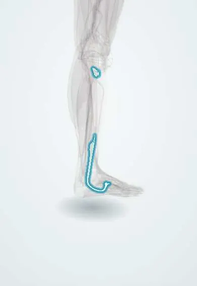
Fibularis longus tendon !
The fibularis longus tendon runs behind the outer ankle bone and under the sole to the big toe's metatarsal; it supports the arch.
Official name: Tendon of fibularis longus
Opens in the Durgical app, no sign-up needed.
What it is
The tendon of the fibularis longus. It runs behind the outer ankle bone, around the cuboid and across the sole to the first metatarsal.
What it does
Turns the foot outward, steadies the ankle and helps support the arches.
Key facts
- Attaches
- Behind the lateral malleolus (superficial to the brevis), below the fibular trochlea, through the groove of the cuboid and across the sole to the medial cuneiform and base of the first metatarsal; an os peroneum may lie in it
- Function
- Eversion and plantarflexion of the first ray; supports the transverse and lateral longitudinal arches
- Tight in
- Inversion with dorsiflexion
Relations
- Course
- Down the lateral leg, behind the lateral malleolus, along the side of the calcaneus, round the groove of the cuboid and obliquely across the deep sole to the inner midfoot
- Deep
- Fibularis brevis tendon behind the malleolus; the calcaneofibular ligament (CFL) below it
- Superficial
- Fibular retinacula; the sural nerve and small saphenous vein run just behind and below
- Below
- In the sole, a tunnel under the cuboid roofed by the long plantar ligament
How to find it
With the foot turned out against resistance, both tendons are felt behind and below the lateral malleolus; the longus is followed under the outer foot toward the cuboid, while the brevis heads to the base of the fifth metatarsal.
Examination
- Resisted eversion: pain or weakness along the tendon suggests tendinopathy or a tear
- Resisted eversion and dorsiflexion while the ankle circles: a tendon felt or seen slipping over the malleolus indicates subluxation
On imaging
On radiographs, an os peroneum that has moved upward or split suggests a longus rupture; dynamic ultrasound shows subluxation; on MRI the curve round the malleolus can show falsely high signal (magic angle effect).
Common problems
- Tendinitis, tears, or slipping out of its groove behind the outer ankle bone, often after ankle sprains.
Clinical notes
- Recurrent dislocation is treated by repairing the retinaculum, sometimes with deepening of the fibular groove
- When one fibular tendon is badly damaged it can be sutured to the other (tenodesis)
- Some centres harvest all or the front half of this tendon as a graft for knee ligament reconstruction; its effect on the arch and the first ray is still debated
Anatomical variants
- Extra slips to the bases of other metatarsals are common; the medial cuneiform attachment is usual but not constant
- An os peroneum is found in a variable share of feet and may be cartilage rather than bone
Memory aid
Behind the lateral malleolus, 'brevis by the bone': the brevis lies against the fibula, the longus outside it.
Test yourself
Think of the answer, then tap the question to see it.
Where does the fibularis longus tendon insert?
On the medial cuneiform and the base of the first metatarsal, after crossing the sole from the groove of the cuboid.
Which sesamoid may lie in the fibularis longus tendon, and why does it matter?
The os peroneum, at the cuboid; a painful, fractured or split os peroneum causes lateral foot pain.
What lets the fibular tendons dislocate over the lateral malleolus, and what is the X-ray sign?
A torn superior fibular retinaculum, usually after forced dorsiflexion; a small flake off the malleolus (fleck sign).
Which fibular tendon lies against the bone behind the lateral malleolus?
The fibularis brevis ('brevis by the bone'); the fibularis longus lies superficial to it.
Educational information, not medical advice. For any symptom, see a health professional.
Its names
- Official Latin (TA2)
- musculus fibularis longus TA2 2652
- Official Spanish (DeCS)
- Tendón del peroneo largo DeCS D013710
- English (TA2)
- Tendon of fibularis longus
- Also called
- musculus peroneus longus · peroneus longus muscle
Related
- CFL
- Long plantar ligament
- Fibular retinacula
- Ankle tendon sheaths
- Fibularis longus
- Cuboid
- Medial cuneiform
- Fibula
Sources: Terminologia Anatomica (TA2); DeCS; Anatomy, Bony Pelvis and Lower Limb: Calf Peroneus Longus Muscle.
Spotted a mistake?We are always improving for the community. Tell us what is wrong and our team will review it.Report a mistake
Representative model, for educational purposes only.