Cardiovascular system · Lower limb
Popliteal artery !
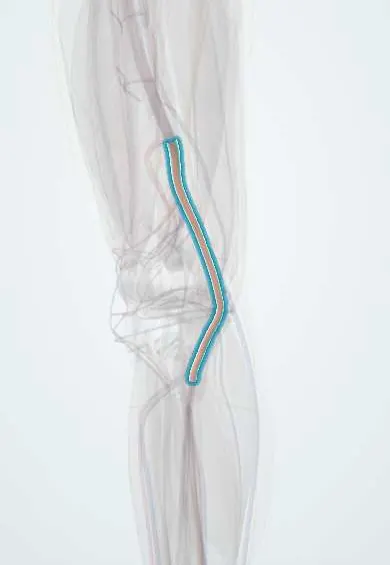
The popliteal artery is the main artery behind the knee, lying deepest in the hollow there; it carries blood to the leg and foot.
Opens in the Durgical app, no sign-up needed.
What it is
The main artery behind the knee, continuing from the femoral artery.
What it does
Carries blood to the lower leg and foot.
Key facts
- Origin
- Continuation of femoral artery at adductor hiatus
- Branches
- Five genicular arteries, sural arteries; ends at lower border of popliteus as anterior and posterior tibial
- Supplies
- Knee joint and calf muscles; leg and foot via terminal branches; deepest structure in popliteal fossa
Relations
- Course
- From the adductor hiatus it runs down and slightly laterally through the popliteal fossa, passes under the fibrous arch of the soleus and divides at the lower border of the popliteus into the anterior tibial artery and posterior tibial artery
- In front
- Popliteal surface of the femur (with a fat pad between), the knee capsule and oblique popliteal ligament, then the fascia over the popliteus
- Behind
- The popliteal vein, bound to it in a common sheath, and the tibial nerve behind that; both cross behind the artery from lateral to medial on the way down
- Medial
- The semimembranosus above, the medial head of gastrocnemius below
- Lateral
- The biceps femoris and lateral femoral condyle above; the lateral head of gastrocnemius and the plantaris below
How to find it
The pulse is deep and easy to miss. With the person supine and the knee partly bent to slacken the popliteal fascia, the examiner wraps both hands around the knee and presses the fingertips into the centre of the fossa against the back of the tibia. An unusually wide, easily felt pulse suggests an aneurysm.
Examination
- Ankle–brachial index with a handheld Doppler after any knee dislocation, compared with the other leg; a value under about 0.9 leads to CT angiography
- Ankle pulses with the knee straight during forced plantar flexion: pulses that fade suggest popliteal entrapment
On imaging
Duplex ultrasound measures an aneurysm and the clot inside it. CT angiography is the quick study after knee trauma and shows the run-off to the foot; catheter angiography allows treatment in the same session. Duplex during plantar flexion, CT or MRI shows entrapment.
Common problems
- Damage in a knee dislocation; this is an emergency.
- Aneurysm (a bulge in the artery wall), more common in older adults.
- Rarely, compression by muscles in young athletes (entrapment).
How it is diagnosed
Checking the pulses in the foot, the ankle-brachial index, ultrasound, CT angiography.
Treatment
- Injury: urgent surgical repair.
- Aneurysm or blockage: monitoring, a bypass or an endovascular repair.
Clinical notes
- Acute ischemia (trauma or embolus) shows the 6 Ps: pain, pallor, pulselessness, paresthesia, paralysis and poikilothermia; nerve and muscle begin to suffer lasting damage within about 6 hours without flow
- Popliteal aneurysm: its main danger is sudden thrombosis or emboli that block the leg arteries, more than rupture
- Popliteal artery entrapment: an abnormal course around the medial head of gastrocnemius squeezes it in young athletes; calf claudication, with ankle pulses that fade on forced plantar flexion
- In knee replacement it lies just behind the posterior capsule, at risk from saws and retractors
- When flow is restored after a long ischaemia, swelling of the leg compartments can cause compartment syndrome
Anatomical variants
- In a large angiographic series (1,242 limbs) about 89% had the usual branching; hypoplastic or absent posterior tibial artery in about 5%, with the fibular artery supplying the sole
- Hypoplastic anterior tibial artery about 1.7%; true trifurcation about 1.5%; high origin of the anterior tibial artery (at or above the knee joint) about 1.2%
- When one limb has a variant, the other has one too in about 28%
Development
Its deep segment comes from the embryonic axial artery. Entrapment results from an abnormal relationship between the developing artery and the medial head of gastrocnemius as that muscle migrates.
Related conditions
Test yourself
Think of the answer, then tap the question to see it.
Which is the deepest of the main structures in the popliteal fossa?
The popliteal artery, lying against the femur and knee capsule; the popliteal vein is superficial to it and the tibial nerve most superficial.
Where does the popliteal artery end, and into which branches?
At the lower border of the popliteus, into the anterior and posterior tibial arteries.
Why does every dislocated knee need a vascular check?
The popliteal artery is fixed at the adductor hiatus and the soleus arch, so a knee dislocation stretches or tears it; pulses and ankle–brachial index are checked even after reduction.
What is the most common peripheral artery aneurysm, and what is its main danger?
Popliteal aneurysm, often bilateral and with an abdominal aortic aneurysm; its danger is thrombosis or emboli rather than rupture.
What are the 6 Ps of acute limb ischemia?
Pain, pallor, pulselessness, paresthesia, paralysis and poikilothermia.
Educational information, not medical advice. For any symptom, see a health professional.
Its names
- Official Latin (TA2)
- arteria poplitea TA2 4699
- Official Spanish (DeCS)
- Arteria poplítea DeCS D011150
- English (TA2)
- Popliteal artery
Related
- Popliteal vein
- Tibial nerve
- Femoral artery
- Genicular arteries
- Gastrocnemius (inner head)
- Gastrocnemius (outer head)
- Plantaris
- Popliteus
Sources: Terminologia Anatomica (TA2); DeCS; Anatomy, Bony Pelvis and Lower Limb: Popliteal Artery; 20.5 Circulatory Pathways (OpenStax (Rice University), 2022) · adapted, CC BY 4.0; Anatomía humana (4.ª ed.), tomo 1, cap. 67: Arterias del miembro inferior.
Spotted a mistake?We are always improving for the community. Tell us what is wrong and our team will review it.Report a mistake
Representative model, for educational purposes only.