Cardiovascular system · Lower limb
Popliteal vein !
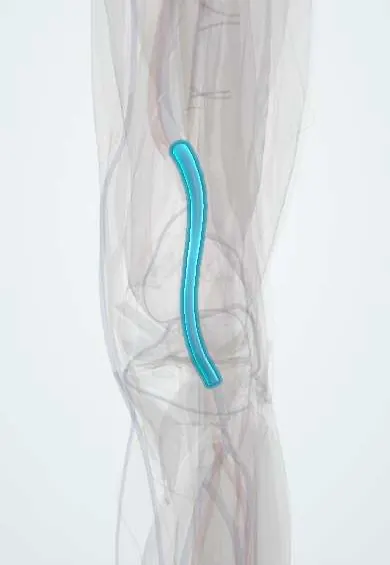
The popliteal vein is the main vein behind the knee, beside the artery; it carries blood from the leg back toward the heart.
Opens in the Durgical app, no sign-up needed.
What it is
The main deep vein behind the knee, next to the artery.
What it does
Returns blood from the lower leg toward the heart.
Key facts
- Drains
- Anterior and posterior tibial and fibular veins (unite at popliteus); small saphenous, genicular and sural veins
- Into
- Femoral vein at adductor hiatus; lies between popliteal artery and tibial nerve
Relations
- Course
- Formed near the lower border of the popliteus, it rises through the fossa, crossing behind the popliteal artery from medial to lateral, and passes through the adductor hiatus to become the femoral vein
- Superficial
- The tibial nerve, then the popliteal fascia, which the small saphenous vein pierces to join it
- Deep
- The popliteal artery, which lies between it and the femur and knee capsule; the two share a tight fascial sheath
How to find it
Not palpable. With the person prone or on the side and the knee slightly bent, ultrasound across the popliteal fossa shows it deep to the tibial nerve and superficial to the popliteal artery; it is followed down to where the calf veins join it and up to the adductor hiatus.
Examination
- Doppler augmentation: squeezing the calf normally gives a brisk rise in flow through the vein; a weak or absent rise suggests obstruction between the calf and the probe
On imaging
On ultrasound, with the person prone or on one side and the knee slightly bent, it lies just superficial to the artery in the middle of the fossa. Clot shows as a vein that will not compress, sometimes with echogenic material inside and no colour flow. It can be doubled, so both channels are checked.
Common problems
- Deep vein thrombosis (a blood clot), more likely after surgery, injury or long periods without moving.
How it is diagnosed
Duplex ultrasound.
Treatment
- Blood thinners; compression. After surgery, moving early and sometimes medicine help prevent clots.
Clinical notes
- A large or ruptured Baker's cyst can cause calf pain and swelling that mimic DVT; ultrasound tells the two apart, and a cyst can also press on the vein
- Injured with the artery in knee dislocation and posterior knee surgery; repairing it helps blood leave the leg after arterial repair
Anatomical variants
- May form higher than usual, close to the adductor hiatus
- Often doubled for part of its course
- Usually posterolateral to the artery, but sometimes medial to it
- Persistent sciatic vein (rare)
Related conditions
Test yourself
Think of the answer, then tap the question to see it.
Why does a clot in the popliteal vein matter more than one confined to the calf?
It is a proximal DVT, with a much higher risk of pulmonary embolism.
What is two-point compression ultrasound, and what shows a clot?
It checks the popliteal and common femoral veins; a normal vein collapses completely under the probe, a vein with clot does not.
Which superficial vein joins the popliteal vein, and where?
The small saphenous vein, at the saphenopopliteal junction; reflux there causes varicose veins on the back of the calf.
Which knee lesion can mimic DVT of the popliteal vein?
A large or ruptured Baker's cyst, which causes calf pain and swelling; ultrasound tells the two apart.
Educational information, not medical advice. For any symptom, see a health professional.
Its names
- Official Latin (TA2)
- vena poplitea TA2 5072
- Official Spanish (DeCS)
- Vena poplítea DeCS D011152
- English (TA2)
- Popliteal vein
Related
Sources: Terminologia Anatomica (TA2); DeCS; Anatomy, Bony Pelvis and Lower Limb: Popliteal Vein.
Spotted a mistake?We are always improving for the community. Tell us what is wrong and our team will review it.Report a mistake
Representative model, for educational purposes only.