Cardiovascular system · Lower limb
Leg veins !
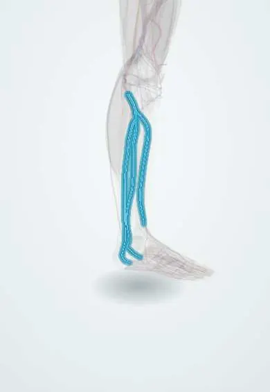
The deep leg veins run with the arteries from the foot up to behind the knee; most deep vein clots begin here.
Official name: Anterior tibial veins
Opens in the Durgical app, no sign-up needed.
What it is
Deep veins that run in pairs with the leg arteries, from the foot up to behind the knee.
What it does
Return blood from the foot and leg to the popliteal vein. Squeezing of the calf muscles helps push the blood up.
Key facts
- Drains
- Foot and leg; paired venae comitantes: anterior tibial, posterior tibial and fibular veins; calf muscle pump
- Into
- Popliteal vein
Relations
- Course
- The posterior tibial veins form behind the medial malleolus from the plantar veins and climb under the soleus; the fibular veins rise along the fibula and join them; the anterior tibial veins climb on the interosseous membrane and pass through the gap above it; all unite near the lower border of the popliteus as the popliteal vein
- Deep
- Deep posterior compartment muscles; the posterior tibial and fibular veins receive the soleal veins, while the gastrocnemius veins mostly drain to the popliteal vein
- Superficial
- Joined to the great saphenous vein system by perforating veins of the medial calf (classically the Cockett perforators), whose valves keep deep pressure away from the skin
How to find it
They cannot be felt. On ultrasound the paired posterior tibial veins flank their artery from behind the medial malleolus up the deep calf, the paired fibular veins lie near the fibula with the fibular artery, and the soleal and gastrocnemius veins sit inside those muscles. The scan usually works down from the popliteal fossa.
Examination
- Wells score: a clinical probability score that, together with the D-dimer, decides who needs an ultrasound for suspected DVT
- Homans sign (calf pain on passive dorsiflexion of the foot) is unreliable and of historical interest only
On imaging
Compression ultrasound shows each paired vein beside its artery. Calf veins are harder to image than the popliteal and femoral veins, so some protocols scan only the proximal veins and repeat the scan about a week later.
Common problems
- Deep vein thrombosis (a blood clot), more likely after a leg injury, surgery or time in a cast.
Clinical notes
- Perforator valve failure sends high pressure to the skin above the medial malleolus (the gaiter area): pigmentation, eczema and venous ulcers
- Immobility, a cast or bed rest stops the calf pump, the basis for early walking and mechanical compression after surgery
Related conditions
Test yourself
Think of the answer, then tap the question to see it.
Where does most DVT start?
In the calf veins: the soleal venous sinuses and the posterior tibial and fibular veins, where blood moves slowest behind the valve cusps.
How does the calf muscle pump return blood?
The soleus and gastrocnemius squeeze the deep veins inside a tight fascial compartment, and valves stop the blood falling back.
What does failure of the calf perforator valves cause?
High pressure on the skin above the medial malleolus (gaiter area): pigmentation, eczema and venous ulcers.
Educational information, not medical advice. For any symptom, see a health professional.
Its names
- Official Latin (TA2)
- venae tibiales anteriores; venae tibiales posteriores; venae fibulares TA2 5075
- Official Spanish (DeCS)
- Venas tibiales anteriores y posteriores y venas fibulares
- English (TA2)
- Anterior tibial veins
- Also called
- peroneal veins
Related
Sources: Terminologia Anatomica (TA2); Anatomy, Bony Pelvis and Lower Limb: Leg Posterior Compartment.
Spotted a mistake?We are always improving for the community. Tell us what is wrong and our team will review it.Report a mistake
Representative model, for educational purposes only.