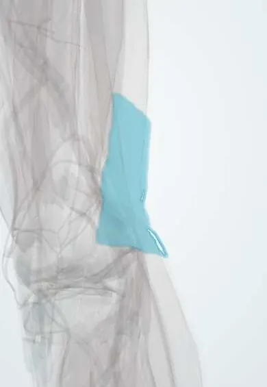
Popliteal fascia !
The popliteal fascia is the sheet roofing the hollow at the back of the knee, covering its nerve, vein and artery.
Opens in the Durgical app, no sign-up needed.
What it is
A sheet covering the hollow at the back of the knee.
What it does
Protects the nerves and vessels behind the knee.
Key facts
- Where
- Roof of popliteal fossa; continuous with fascia lata above and crural fascia below
- Function
- Covers popliteal fossa contents; pierced by small saphenous vein; tight roof makes popliteal swellings painful
Relations
- Superficial
- Skin and subcutaneous fat with the small saphenous vein (before it pierces the fascia) and branches of the posterior femoral cutaneous nerve
- Deep
- Popliteal fossa: tibial nerve and common fibular nerve, popliteal vein and artery, popliteal lymph nodes and fat; the medial sural cutaneous nerve runs just under it
- Above
- Fascia lata of the back of the thigh, over the hamstrings
- Below
- Crural fascia over the two heads of gastrocnemius
How to find it
The popliteal fossa is the diamond-shaped hollow behind the knee. With the knee straight the fascia is taut and the hollow feels firm; with the knee bent and relaxed it slackens, and the popliteal pulse is sought deep in the centre of the fossa, pressed against the bone.
On imaging
Not assessed on its own; it is the roof of the popliteal fossa. Ultrasound and MRI of the fossa look below it at the vessels (artery deepest, vein superficial to it), the tibial nerve and any mass. The common finding is a Baker cyst between the medial gastrocnemius head and the semimembranosus tendon. Duplex ultrasound checks for popliteal aneurysm or vein thrombosis.
Clinical notes
- Posterior approach to the knee: the fascia is opened next to the small saphenous vein, and the medial sural cutaneous nerve under it is followed up to find the tibial nerve
- Swellings bound by the tight fascia (popliteal cyst, aneurysm, tumour) can compress the tibial nerve or popliteal vein before they become visible
- Incisions behind the knee are made S-shaped or zigzag so that they cross the flexion crease obliquely; a straight vertical scar across the crease tends to contract and limit extension
Anatomical variants
- The level at which the small saphenous vein pierces the fascia and ends varies; it may continue up the thigh to the deep veins or join the great saphenous vein (Giacomini vein)
Test yourself
Think of the answer, then tap the question to see it.
Under the popliteal fascia, what is the order of the main neurovascular structures from superficial to deep?
Tibial nerve, popliteal vein, popliteal artery; the artery lies deepest, on the femur and the knee capsule.
What does an unusually easy-to-feel popliteal pulse suggest, given the tension of the popliteal fascia?
A popliteal artery aneurysm: the tight fascia normally makes the popliteal pulse hard to feel.
Which superficial vein usually pierces the popliteal fascia, and where does it end?
The small saphenous vein; it usually ends in the popliteal vein, although the level varies.
Educational information, not medical advice. For any symptom, see a health professional.
Its names
- Official Latin (TA2)
- fascia poplitea TA2 2707
- Official Spanish (DeCS)
- Fascia poplítea DeCS D005205
- English (TA2)
- Popliteal fascia
Sources: Terminologia Anatomica (TA2); DeCS; Anatomy, Bony Pelvis and Lower Limb: Popliteal Region.
Spotted a mistake?We are always improving for the community. Tell us what is wrong and our team will review it.Report a mistake
Representative model, for educational purposes only.