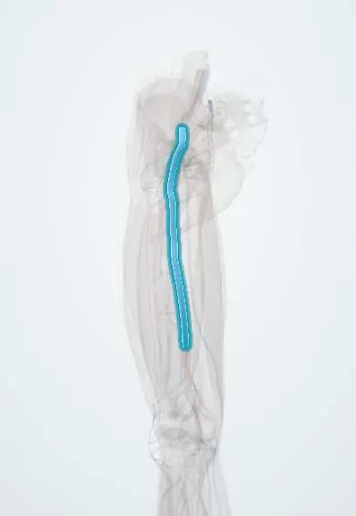
Deep vein thrombosis !
Deep vein thrombosis is a blood clot in a deep vein, usually of the leg; it can cause a swollen, painful leg and can travel to the lungs.
Updated 2026-10-30
Structures involved
Tap one to see what it is and open it in 3D.
- Femoral vein
- Popliteal vein
- Leg veins
- Deep femoral vein
- External iliac vein
- Great saphenous vein
- Small saphenous vein
- Soleus
- Gastrocnemius (inner head)
In biochemistry
In histology
In embryology
In the clinical exam
- Signs of deep vein thrombosis: Homans and the Wells score
- Varicose veins: the Trendelenburg (Brodie–Trendelenburg) test
- Edema: pitting, grading and distribution
In physiology
- The coagulation cascade
- Fibrinolysis and natural anticoagulants
- Thrombosis and bleeding: Virchow's triad and hemostatic defects
Why it happens
- Hypercoagulable states: inherited and acquired
- Thrombosis and Virchow's triad
- Arterial vs venous thrombi; mural thrombi and lines of Zahn
- What happens to a thrombus: propagation, embolism, dissolution, organization
- Pulmonary thromboembolism: where it comes from and what it does
What it is
A blood clot (thrombus) in a deep vein, most often of the leg or pelvis. Together with pulmonary embolism it forms venous thromboembolism.
Who gets it
Common in hospitalised, post-surgical, trauma and cancer patients, and more frequent with age. Latin American data are scarce: a 2016 review cites an Argentine estimate, extrapolated from one hospital, of 0.48 DVT cases per 1,000 person-years, and about half of hospitalised patients in Mexico and Brazil were at risk. Thromboprophylaxis is standard hospital care.
Causes
- Virchow's triad: slow or stagnant flow, vein-wall injury and a hypercoagulable state
- Immobility after surgery, trauma, plaster casts or long journeys
- Inherited or acquired clotting disorders
Risk factors
- Recent major surgery, especially hip or knee surgery, or hip fracture
- Active cancer
- Previous venous thromboembolism
- Pregnancy, the postpartum period, and oestrogen-containing contraception or hormone therapy
- Older age, obesity and prolonged bed rest
- Central venous catheters
Pathophysiology
Clots usually start in the valve pockets of calf veins where flow is slow. They can grow upward into the popliteal, femoral and iliac veins (proximal DVT). Pieces of a proximal clot can break off and lodge in the pulmonary arteries. Damage to the valves can leave lasting venous hypertension.
Classification
- Distal (calf veins only)
- Proximal (popliteal vein and above)
- Provoked (clear transient trigger) or unprovoked
Symptoms and signs
- Swelling of one leg, often with a calf circumference difference
- Pain or tenderness along the deep veins
- Warmth and redness of the leg
- Prominent superficial veins (non-varicose)
- May be silent: many clots cause few or no signs
Diagnosis
- Clinical
- Pre-test probability with the Wells score for DVT guides the work-up; signs alone are unreliable.
- Tests
- D-dimer: a normal result helps exclude DVT when probability is low; it is non-specific (raised after surgery, in pregnancy, cancer, infection). Baseline blood count, clotting tests and kidney function before treatment.
- Imaging
- Compression ultrasound (a vein that does not collapse under the probe holds a clot) is the test of choice. CT or MR venography for pelvic veins.
Differential diagnosis
- Baker's cyst rupture
- Cellulitis
- Calf muscle strain or tear
- Superficial thrombophlebitis
- Lymphoedema or heart, liver or kidney oedema
Goal of treatment
Stop the clot from growing, prevent pulmonary embolism and recurrence, and limit post-thrombotic syndrome, while keeping bleeding risk low.
Treatment
- Medical
- Anticoagulation is the mainstay: low-molecular-weight heparin, unfractionated heparin, direct oral anticoagulants or vitamin K antagonists, chosen by the patient's profile; length depends on provoked or unprovoked cause.
- Procedure
- Thrombolysis or catheter-directed clot removal for selected cases (e.g. limb-threatening DVT); an inferior vena cava filter when anticoagulation cannot be given.
- Conservative
- Early walking once anticoagulated; compression stockings may help symptoms, though their role in prevention is debated.
- Follow-up
- Follow-up for bleeding, recurrence and post-thrombotic symptoms; cancer screening is considered in some unprovoked cases.
Drugs for this condition
Complications
- Pulmonary embolism, which can be fatal
- Post-thrombotic syndrome (chronic swelling, pain, skin changes, ulcers)
- Recurrent venous thromboembolism
- Bleeding from anticoagulation
- Chronic thromboembolic pulmonary hypertension (after PE)
Prognosis
With prompt anticoagulation most patients recover well. Recurrence is more likely after unprovoked clots, and a proportion develop post-thrombotic syndrome.
Prevention
- Risk assessment and thromboprophylaxis (drug or mechanical) for hospitalised and surgical patients
- Early mobilisation after surgery
- Moving the legs and staying hydrated on long journeys
Test yourself
Think of the answer, then tap the question to see it.
Which three elements make up Virchow's triad?
Venous stasis, endothelial (vein-wall) injury and hypercoagulability.
What is the first-line imaging test for suspected leg DVT?
Compression ultrasound: a deep vein that cannot be compressed contains a thrombus.
When is a normal D-dimer most useful?
When clinical probability (e.g. Wells score) is low: a normal D-dimer then helps rule out DVT. A raised D-dimer is non-specific.
What is the most feared acute complication of proximal DVT?
Pulmonary embolism, when part of the clot travels to the pulmonary arteries.
What chronic complication causes leg swelling, skin changes and ulcers after DVT?
Post-thrombotic syndrome, from valve damage and persistent venous hypertension.
Related conditions
- Intertrochanteric hip fracture
- Femoral Neck Fracture
- Varicose veins
- Baker's (Popliteal) Cyst
- Lymphoedema
- Patellar fracture
- Ankle fracture
- Achilles tendon rupture
Educational information, not medical advice. For any symptom, see a health professional.
Sources: Deep Venous Thrombosis (StatPearls (NCBI Bookshelf), 2023); Venous thromboembolism in Latin America: a review and guide to diagnosis and treatment for primary care (Clinics (Sao Paulo, Brazil), 2016) · adapted, CC BY 4.0.
Spotted a mistake?We are always improving for the community. Tell us what is wrong and our team will review it.Report a mistake
Representative model, for educational purposes only.