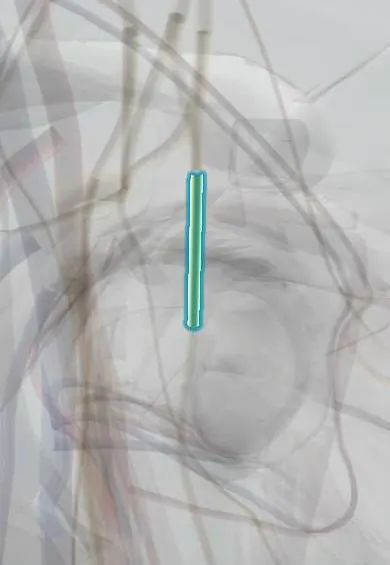
Posterior cord !
The posterior cord of the brachial plexus lies in the armpit behind the axillary artery; it gives the axillary and radial nerves.
Official name: Posterior cord of the brachial plexus
Opens in the Durgical app, no sign-up needed.
What it is
The back bundle of the plexus, behind the axillary artery.
What it does
Gives the axillary and radial nerves.
Key facts
- Roots
- C5–T1 (all posterior divisions)
- Moves
- Upper and lower subscapular, thoracodorsal, axillary and radial nerves
- Feeling from
- Via the axillary and radial nerves
Relations
- Course
- Formed behind the first part of the axillary artery by the three posterior divisions, it lies behind the second part, deep to pectoralis minor, and ends at the muscle's lower border by dividing into the axillary nerve and the radial nerve
- In front
- The axillary artery (second part), with pectoralis minor in front of it
- Behind
- Subscapularis and the posterior wall of the axilla, down which its subscapular and thoracodorsal branches run
How to find it
In a dissection, lifting the axillary artery forward below the clavicle shows the posterior cord directly behind it on subscapularis. On infraclavicular ultrasound it lies just deep to the artery, the usual target for a single injection that spreads to all three cords.
Examination
- Deltoid, latissimus dorsi (cough test on the posterior axillary fold), triceps, and wrist and finger extensors: weakness across axillary, thoracodorsal and radial muscles places the lesion at the cord
- Triceps reflex (C7) reduced; pinprick over the lower deltoid and the dorsum of the first web space
On imaging
On infraclavicular ultrasound and sagittal MRI it lies behind the second part of the axillary artery, the deepest of the three cords. MRI is the main test after trauma, looking for swelling, discontinuity or scar. With an anterior shoulder dislocation or proximal humerus fracture, radiographs show the cause and its axillary and radial branches are checked clinically.
Common problems
- Stretch injuries from motorcycle crashes or falls that force the head and shoulder apart.
- Birth injury (Erb's palsy) affecting the upper trunk: the arm hangs turned inward ('waiter's tip').
- Compression at the thoracic outlet between the collarbone and the first rib.
How it is diagnosed
A detailed exam of strength and feeling in each nerve's area. MRI, EMG and nerve conduction studies show the level and severity.
Treatment
- Physical therapy and watching for recovery; many stretch injuries improve.
- Nerve repair, graft or nerve transfer surgery for injuries that do not recover, ideally within months.
Clinical notes
- Posterior cord lesions follow anterior shoulder dislocation, proximal humerus fractures and penetrating axillary wounds: deltoid and wrist extensor weakness with a normal biceps
- Crutch palsy vs Saturday night palsy: crutch pressure in the axilla hits the radial nerve above its triceps branches; an arm hung over a chair compresses it in the spiral groove and spares the triceps
- Posterior cord vs C7 radiculopathy: C7 also weakens wrist flexion and pronation (flexor carpi radialis, pronator teres) and spares the deltoid; a cord lesion does the opposite
Anatomical variants
- Prefixed and postfixed plexuses change its root make-up, and some of its branches (the thoracodorsal or lower subscapular nerve) may arise from the axillary or radial nerve instead of the cord itself
Development
It gathers all the posterior divisions, the nerves of the dorsal (extensor) muscle mass. When the upper limb turns outward by about 90° in week 7, that mass ends up facing backward, so every muscle the cord supplies lies on the back of the shoulder, arm or forearm, or in the posterior axillary wall.
Memory aid
Posterior cord branches, 'ULTRA': Upper subscapular, Lower subscapular, Thoracodorsal, Radial, Axillary.
Related conditions
Test yourself
Think of the answer, then tap the question to see it.
How is the posterior cord of the brachial plexus formed, and which branches does it give?
By all three posterior divisions (C5–T1); it gives the upper and lower subscapular, thoracodorsal, axillary and radial nerves.
How does crutch palsy differ from Saturday night palsy?
Crutch pressure in the axilla hits the posterior cord or high radial nerve, so the triceps is weak; an arm over a chair compresses the spiral groove and spares the triceps.
How is a posterior cord lesion told apart from a C7 radiculopathy?
C7 also weakens wrist flexion and pronation and spares the deltoid; a posterior cord lesion weakens the deltoid and spares those.
Educational information, not medical advice. For any symptom, see a health professional.
Its names
- Official Latin (TA2)
- fasciculus posterior plexus brachialis TA2 6416
- Official Spanish (DeCS)
- Fascículo posterior del plexo braquial
- English (TA2)
- Posterior cord of the brachial plexus
Related
Sources: Terminologia Anatomica (TA2); Anatomy, Shoulder and Upper Limb, Brachial Plexus; Anatomy, Head and Neck: Brachial Plexus; 8.5 Development of the Appendicular Skeleton (OpenStax (Rice University), 2022) · adapted, CC BY 4.0; Anatomy of the Human Body (20th ed.): The Branchial Region (Lea & Febiger (Gray's Anatomy, 1918), 1918) · adapted, Public domain.
Spotted a mistake?We are always improving for the community. Tell us what is wrong and our team will review it.Report a mistake
Representative model, for educational purposes only.