Cardiovascular system · Head and neck
Retromandibular vein !
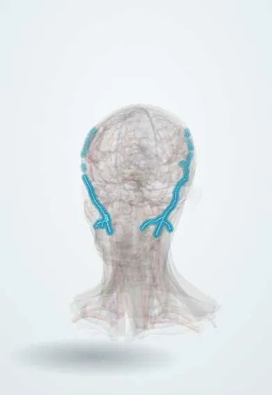
The retromandibular vein runs behind the jaw through the parotid gland, draining the temple and deep face; it guides surgeons in parotid surgery.
Opens in the Durgical app, no sign-up needed.
What it is
A vein behind the jaw inside the parotid gland.
What it does
Drains the temple and the deep face; a landmark in parotid surgery.
Key facts
- Drains
- Superficial temporal and maxillary veins (pterygoid plexus)
- Into
- Anterior division -> facial vein; posterior division -> external jugular
Relations
- Course
- Forms in the parotid gland behind the neck of the mandible, runs down through the gland behind the ramus and divides near the gland's lower pole
- Superficial
- Facial nerve branches within the parotid and the superficial lobe of the gland
- Deep
- External carotid artery, which ends in the parotid by dividing into the superficial temporal and maxillary arteries
- In front
- Ramus of the mandible and the masseter
How to find it
It cannot be felt. It runs down behind the ramus of the mandible inside the parotid gland; on ultrasound and CT it is seen within the gland, and in parotid surgery it is met at the lower pole, where the marginal mandibular branch of the facial nerve is often found crossing it.
On imaging
On axial contrast CT or MRI it is a small round vessel in the parotid just behind the ramus. A mass lateral to it is usually in the superficial lobe; one medial to it is in the deep lobe and may extend into the parapharyngeal space.
Clinical notes
- A source of bleeding when the superficial lobe of the parotid is lifted; the facial nerve is traced where it crosses the vein
- Its divisions guide the surgeon down to the facial vein and the external jugular vein at the lower pole of the gland
Anatomical variants
- Its divisions vary: it may not divide and drain entirely to the facial vein or to the external jugular vein
- In a minority of people the facial nerve, or one of its branches, passes deep to the vein
Development
It begins as part of the embryonic linguofacial drainage; cross-links that form later give part of its blood to the external jugular, which is why it usually splits into anterior and posterior divisions.
Related conditions
Test yourself
Think of the answer, then tap the question to see it.
What is the order of structures in the parotid gland, from superficial to deep?
Facial nerve branches, then the retromandibular vein, then the external carotid artery.
How is the retromandibular vein used on CT and MRI of the parotid?
As the line splitting the gland into superficial and deep lobes, because the facial nerve itself is not seen.
Where do the divisions of the retromandibular vein drain?
Anterior division to the facial vein; posterior division to the external jugular vein.
Educational information, not medical advice. For any symptom, see a health professional.
Its names
- Official Latin (TA2)
- vena retromandibularis TA2 4831
- Official Spanish (DeCS)
- Vena retromandibular
- English (TA2)
- Retromandibular vein
- Also called
- vena facialis posterior · posterior facial vein
Related
Sources: Terminologia Anatomica (TA2); Anatomy, Head and Neck, Parotid Gland; Anatomy, Head and Neck, External Jugular Veins; Anatomy of the Human Body (20th ed.): Development of the Vascular System (Lea & Febiger (Gray's Anatomy, 1918), 1918) · adapted, Public domain.
Spotted a mistake?We are always improving for the community. Tell us what is wrong and our team will review it.Report a mistake
Representative model, for educational purposes only.