Nervous system · Head and neck
Facial nerve branches !
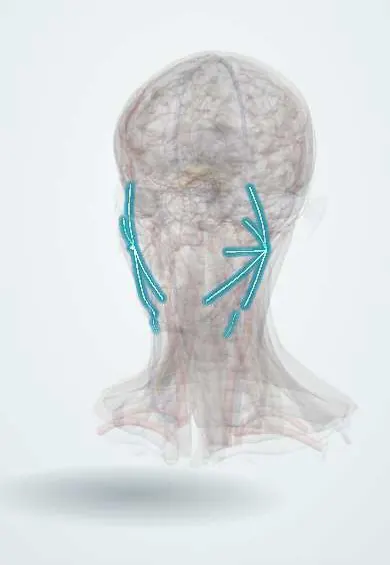
The five branches of the facial nerve fan out from the parotid gland across the face; each moves one zone, from the forehead to the neck.
Official name: Temporal branches of facial nerve
Opens in the Durgical app, no sign-up needed.
What it is
The five branches of the facial nerve after it passes through the parotid gland.
What it does
Each moves one zone of the face; surgeons map them in parotid and facelift surgery.
Key facts
- Roots
- Facial nerve after the parotid: temporal, zygomatic, buccal, marginal mandibular, cervical
- Moves
- Each supplies one zone of the facial muscles
- Feeling from
- None
Relations
- Course
- Emerge from the upper, front and lower borders of the parotid gland and fan out across the face toward the temple, cheekbone, cheek, jaw line and neck
- Superficial
- Superficial musculoaponeurotic system (SMAS) and platysma, under the skin and fat
- Deep
- Masseter and the buccal fat; most branches enter the facial muscles from their deep surface (except buccinator, mentalis and levator anguli oris)
- Below
- The marginal mandibular branch runs along the lower border of the mandible, crossing over the facial artery and vein; the cervical branch descends to the platysma
How to find it
The temporal branch crosses the zygomatic arch roughly along a line from just below the tragus to just above the lateral end of the eyebrow. Buccal branches run with the parotid duct across the masseter. The marginal mandibular branch crosses the facial vessels where the facial artery pulse is felt at the front edge of the masseter.
Examination
- Temporal: raise the eyebrows and wrinkle the forehead
- Zygomatic: close the eyes tightly against resistance
- Buccal: smile, puff the cheeks, whistle
- Marginal mandibular: show the lower teeth or push the lower lip out
- Cervical: tense the neck skin by pulling the corners of the mouth down
On imaging
The branches are not seen on routine imaging, so their plane is inferred: the retromandibular vein, seen on ultrasound, CT or MRI, runs just deep to the nerve and marks the line between the superficial and deep parts of the parotid. Placing a tumour on either side of it helps plan surgery. Ultrasound guides needle biopsy; MRI shows deep extension and perineural spread.
Clinical notes
- Parotidectomy dissects along the nerve from the trunk out to the branches, often with nerve monitoring
- Lacerations lateral to a vertical line through the lateral canthus may cut major branches and are explored and repaired; medial to it, small branches usually recover
- Neck incisions are placed well below the lower border of the mandible to protect the marginal mandibular branch
- Zygomatic and buccal branches interconnect widely, so isolated injuries often recover
Anatomical variants
- The branching pattern within the parotid varies widely, with many described types and frequent links between the zygomatic and buccal branches
Development
The facial muscles come from the second (hyoid) arch, and the facial nerve spreads with them over the face. At birth there is no mastoid process, so the trunk leaves close to the surface below the ear and can be hurt by forceps or by an incision behind the ear in an infant.
Memory aid
«To Zanzibar By Motor Car»: Temporal, Zygomatic, Buccal, Marginal mandibular, Cervical, from top to bottom.
Related conditions
Test yourself
Think of the answer, then tap the question to see it.
What are the five terminal branches of the facial nerve, from top to bottom?
Temporal, zygomatic, buccal, marginal mandibular and cervical, fanning out from the parotid gland.
Which facial nerve branches are most likely to be permanently injured, and why?
The temporal and marginal mandibular branches: they have few connections with the other branches.
What does injury to the marginal mandibular branch of the facial nerve cause, and how is it protected?
Weak lower lip depressors and an asymmetric smile; neck incisions are placed well below the lower border of the mandible.
Which facial lacerations are explored for repair of the facial nerve branches?
Those lateral to a vertical line through the lateral canthus, which may cut major branches; medial to it, small branches usually recover.
Educational information, not medical advice. For any symptom, see a health professional.
Its names
- Official Latin (TA2)
- rami temporales nervi facialis TA2 6302
- Official Spanish (DeCS)
- Ramas del nervio facial
- English (TA2)
- Temporal branches of facial nerve
- Also called
- rami temporofrontales
Sources: Terminologia Anatomica (TA2); Facial Nerve Anatomy and Clinical Applications; Bell Palsy; Anatomy of the Human Body (20th ed.): Development of the Muscles (Lea & Febiger (Gray's Anatomy, 1918), 1918) · adapted, Public domain.
Spotted a mistake?We are always improving for the community. Tell us what is wrong and our team will review it.Report a mistake
Representative model, for educational purposes only.