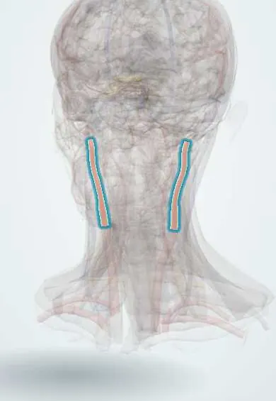
Cardiovascular system · Head and neck
External carotid artery !
The external carotid artery supplies the outside of the head: the thyroid, tongue, face, scalp and jaws, through branches in the neck.
Opens in the Durgical app, no sign-up needed.
What it is
The branch that supplies the face, scalp, neck and throat.
What it does
Gives the thyroid, tongue, face, scalp and jaw arteries.
Key facts
- Origin
- Carotid bifurcation
- Branches
- Superior thyroid, ascending pharyngeal, lingual, facial, occipital, posterior auricular, maxillary, superficial temporal
- Supplies
- Face, scalp, neck, thyroid, tongue, jaws, dura (middle meningeal)
Relations
- Course
- From the bifurcation at the upper border of the thyroid cartilage it rises in the carotid triangle, passes deep to the posterior belly of the digastric and the stylohyoid, enters the parotid gland and divides behind the neck of the mandible into the maxillary artery and the superficial temporal artery
- Superficial
- Sternocleidomastoid at its start, then the hypoglossal nerve, the facial and lingual veins, the digastric and stylohyoid; in the parotid gland, the retromandibular vein and the facial nerve lie superficial to it
- Deep
- Pharyngeal wall and the superior laryngeal nerve; higher up, the styloglossus, stylopharyngeus, glossopharyngeal nerve and pharyngeal branch of the vagus nerve pass between it and the internal carotid artery
How to find it
It is not felt apart from the carotid pulse, but its branches are: the facial artery on the lower border of the mandible and the superficial temporal artery in front of the tragus. In neck dissection it is identified by its branches, usually the superior thyroid artery first, and by the hypoglossal nerve crossing it.
Examination
- Temporal tap on duplex ultrasound: tapping the superficial temporal artery in front of the ear sends ripples into the external carotid waveform, which helps tell it from the internal carotid
On imaging
Duplex ultrasound: branches in the neck, a high-resistance waveform with little flow in diastole, and small ripples in the trace when the superficial temporal artery is tapped in front of the ear (temporal tap).
Clinical notes
- Ligation or embolisation of the external carotid or its branches is used for bleeding from the nose, tongue or face that local measures do not stop
- When the internal carotid is blocked, external carotid branches can feed the brain through the orbit (facial and superficial temporal links with the ophthalmic artery)
- Those same links are 'dangerous anastomoses' during embolisation: particles can reach the eye or the brain
- The superior thyroid artery runs close to the external laryngeal nerve; tying it high in a thyroidectomy can injure the nerve and weaken the high-pitched voice
- It is found for ligation in Farabeuf's triangle (internal jugular vein behind, the common facial venous trunk below, the hypoglossal nerve above), and is identified by its branches before tying, so the internal carotid is not tied by mistake
Anatomical variants
- The lingual and facial arteries often arise from a common linguofacial trunk; a thyrolingual trunk is less common
- The superior thyroid artery sometimes arises from the common carotid artery or at the bifurcation
Development
It grows from the ventral aorta (some texts: from a bud of the third arch artery), while the internal carotid forms from the third arch artery and the dorsal aorta.
Memory aid
Branches from below up: "Some Anatomists Like Freaking Out Poor Medical Students" (Superior thyroid, Ascending pharyngeal, Lingual, Facial, Occipital, Posterior auricular, Maxillary, Superficial temporal).
Related conditions
Test yourself
Think of the answer, then tap the question to see it.
How is the external carotid artery told apart from the internal carotid in the neck?
It has branches in the neck (usually the superior thyroid first) and starts anteromedial to the internal carotid.
What are the branches of the external carotid artery, from below up?
Superior thyroid, ascending pharyngeal, lingual, facial, occipital, posterior auricular, maxillary and superficial temporal.
Why can the external carotid artery be tied off to stop bleeding without losing the face or scalp?
Its branches anastomose freely across the midline and with internal carotid branches around the orbit.
Where does the external carotid artery end?
In the parotid gland, behind the neck of the mandible, dividing into the maxillary and superficial temporal arteries.
Educational information, not medical advice. For any symptom, see a health professional.
Its names
- Official Latin (TA2)
- carotis externa TA2 4369
- Official Spanish (DeCS)
- Arteria Carótida Externa DeCS D002342
- English (TA2)
- External carotid artery
Related
Sources: Terminologia Anatomica (TA2); DeCS; Anatomy, Head and Neck: Carotid Arteries; Anatomy of the Human Body (20th ed.): Development of the Vascular System (Lea & Febiger (Gray's Anatomy, 1918), 1918) · adapted, Public domain; Tratado de anatomía humana (9.ª ed.), tomo II, Libro IV, sección 2 Arterias, caps. I-III (sistema de la arteria pulmonar; aorta y ramas del cayado hasta la carótida interna).
Spotted a mistake?We are always improving for the community. Tell us what is wrong and our team will review it.Report a mistake
Representative model, for educational purposes only.