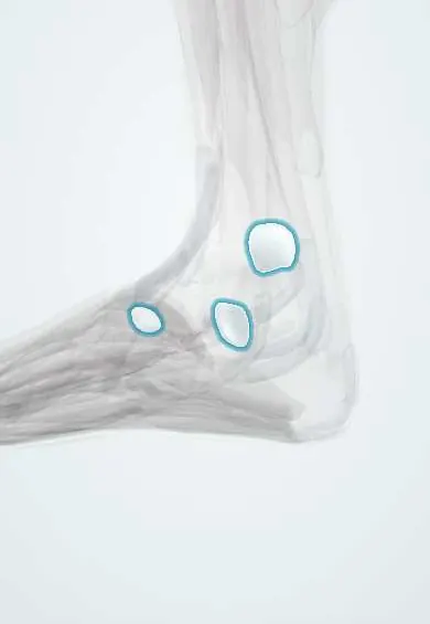
Ankle bursae !
The ankle bursae are small fluid sacs over the ankle bones and under the front shin tendon; they reduce rubbing.
Official name: Subcutaneous bursa of lateral malleolus
Opens in the Durgical app, no sign-up needed.
What it is
Small fluid sacs under the skin over the inner and outer ankle bones, and under the tibialis anterior tendon.
What it does
Reduce friction where skin or tendons move over bone.
Key facts
- Where
- Subcutaneous bursae over medial and lateral malleoli; subtendinous bursa of tibialis anterior
- Function
- Reduce friction of skin and tendons over bony points
Relations
- Superficial
- Skin and thin fat; the great saphenous vein and saphenous nerve pass in front of the medial malleolus, the small saphenous vein and sural nerve behind the lateral one
- Deep
- Subcutaneous surfaces of the medial and lateral malleoli; for the tibialis anterior bursa, the medial cuneiform under the tendon
How to find it
Both malleoli lie just under the skin. A malleolar bursa is a well-defined swelling over the bony tip that moves with the skin; tendon sheath swelling follows a tendon behind or below the malleolus, and an ankle effusion fills the front of the joint.
On imaging
Ultrasound shows a compressible fluid collection between the skin and the malleolus, separate from the tendon sheaths and the joint. MRI is kept for swellings that look solid or atypical.
Common problems
- Can swell from pressure, such as tight boots or sitting cross-legged.
Clinical notes
- The differential of a lump over the malleolus includes bursitis, tenosynovitis, an ankle effusion, a ganglion and a lipoma; examination and ultrasound usually settle it
- Subcutaneous malleolar bursae are often acquired, forming over the lateral malleolus in people who sit cross-legged or on the floor and under tight boots or casts
- A red, hot, tender malleolar bursa may be septic or crystal-related (gout); aspirated fluid is sent for cell count, Gram stain, culture and crystals
Related conditions
Test yourself
Think of the answer, then tap the question to see it.
Which bursae make up the ankle bursae?
Subcutaneous bursae over the medial and lateral malleoli and the subtendinous bursa of the tibialis anterior.
How does malleolar bursitis differ from an ankle effusion on examination?
It is a soft swelling directly over the bone, and the ankle moves freely and painlessly; an effusion fills the front of the joint.
Why is a malleolar pressure bursa a concern in diabetic neuropathy?
It can ulcerate and become infected.
Educational information, not medical advice. For any symptom, see a health professional.
Its names
- Official Latin (TA2)
- bursa subcutanea malleoli lateralis; bursa subcutanea malleoli medialis TA2 2749
- Official Spanish (DeCS)
- Bolsas subcutáneas de los maléolos DeCS D002061
- English (TA2)
- Subcutaneous bursa of lateral malleolus
- Also called
- bursa subcutanea malleoli fibulae
Related
Sources: Terminologia Anatomica (TA2); DeCS; Anatomy, Skin Bursa.
Spotted a mistake?We are always improving for the community. Tell us what is wrong and our team will review it.Report a mistake
Representative model, for educational purposes only.