Cardiovascular system · Lower limb
Great saphenous vein !
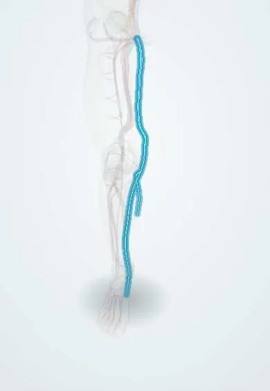
The great saphenous vein is a long surface vein from the inner ankle up to the groin, where it joins the femoral vein.
Opens in the Durgical app, no sign-up needed.
What it is
The longest vein in the body: a surface vein running from the inner ankle up the inner leg and thigh to the groin, where it joins the femoral vein.
What it does
Drains blood from the skin and fat of the leg. One-way valves keep the blood flowing upward.
Key facts
- Drains
- Medial end of dorsal venous arch, anterior to medial malleolus; superficial medial leg and thigh
- Into
- Femoral vein through saphenous opening, below and lateral to pubic tubercle (saphenofemoral junction)
Relations
- Course
- From the medial end of the dorsal venous arch it passes in front of the medial malleolus, climbs the medial leg with the saphenous nerve, passes about a hand's breadth behind the medial border of the patella, ascends the medial thigh and pierces the cribriform fascia to join the femoral vein
- Above
- At the junction it receives the superficial epigastric, superficial circumflex iliac and superficial external pudendal veins, often with anterior and posterior accessory saphenous veins
- Deep
- The deep fascia (fascia lata in the thigh); perforating veins (classically Cockett in the lower leg, Boyd below the knee, Dodd and Hunter in the thigh) link it or its tributaries to the deep veins; at the junction the deep external pudendal artery passes between the arch and the femoral vein
- Lateral
- At the groin, the femoral artery pulse, a little lateral to the junction
How to find it
With the person standing so the veins fill, it is seen and felt as a soft cord just in front of the medial malleolus and along the inner side of the lower leg. In the thigh it is usually visible only when varicose. Its junction lies in the groin just medial to the femoral pulse.
Examination
- Trendelenburg (tourniquet) test: the leg is raised to empty the veins and the junction is compressed; if the veins fill quickly from above when the pressure is released on standing, the saphenofemoral valve is incompetent (largely replaced by duplex ultrasound)
- Cough impulse at the junction: a soft groin swelling that fills on standing, gives a thrill on coughing and empties on lying down is a saphena varix, a mimic of femoral hernia
On imaging
Duplex ultrasound with the person standing maps reflux: backflow lasting more than about half a second after a calf squeeze is abnormal. In the thigh the vein sits in its own fascial compartment, seen in cross-section as the «saphenous eye».
Common problems
- Varicose veins, when its valves leak.
- Superficial thrombophlebitis: a painful, inflamed vein with a clot near the surface.
How it is diagnosed
Physical exam; duplex ultrasound to check for leaking valves and clots.
Treatment
- Compression stockings, raising the legs and staying active.
- Closing the vein with heat (laser or radiofrequency) or other methods, or removing it, for troublesome varicose veins.
- Healthy segments are often used as a graft in heart or leg bypass surgery.
Clinical notes
- Thermal or chemical ablation from inside the vein has largely replaced surgical stripping for incompetent trunks
- Superficial thrombophlebitis close to the saphenofemoral junction can extend into the femoral vein, which is why it is studied with ultrasound
- Two valves guard the junction: the terminal valve at the femoral vein and a preterminal valve a few centimetres lower, between which the junction tributaries enter; duplex scanning checks both before treatment
Anatomical variants
- Accessory saphenous veins running parallel to it in the thigh are common
- The main channel may leave its fascial compartment for part of the thigh and run just under the skin, where a parallel tributary can be mistaken for it
Development
It forms from the preaxial (medial) marginal vein of the limb bud, while the small saphenous vein comes from the postaxial (lateral) one; this is why one starts at the medial end and the other at the lateral end of the dorsal venous arch.
Related conditions
- Varicose veins
- Chronic venous insufficiency
- Venous leg ulcer
- Inherited thrombophilia
- Lymphatic filariasis
- Superficial thrombophlebitis (superficial vein thrombosis)
Test yourself
Think of the answer, then tap the question to see it.
Where is the great saphenous vein constant, making it the classic site for an emergency venous cutdown?
In front of the medial malleolus.
Where is the saphenofemoral junction, and what does failure of its valve cause?
A few centimetres (classically about 3–4 cm) below and lateral to the pubic tubercle; failure of its terminal valve is the commonest start of varicose veins.
Which nerve runs with the great saphenous vein below the knee?
The saphenous nerve, so stripping or harvesting the lower-leg segment can leave numbness of the medial leg and foot.
Why is the great saphenous vein reversed, or its valves cut, when used as a bypass conduit?
So its valves do not block arterial flow; it is the most used conduit for coronary and leg bypass.
Educational information, not medical advice. For any symptom, see a health professional.
Its names
- Official Latin (TA2)
- vena saphena magna TA2 5058
- Official Spanish (DeCS)
- Vena safena mayor DeCS D012501
- English (TA2)
- Great saphenous vein
- Also called
- long saphenous vein
Related
Sources: Terminologia Anatomica (TA2); DeCS; Anatomy, Bony Pelvis and Lower Limb: Saphenous Nerve, Artery, and Vein; Tratado de anatomía humana (9.ª ed.), tomo II, Libro IV Angiología, sección 4: Venas (sistemas de las venas cavas superior e inferior, vena porta, venas del raquis).
Spotted a mistake?We are always improving for the community. Tell us what is wrong and our team will review it.Report a mistake
Representative model, for educational purposes only.