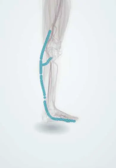
Sural nerves !
The sural nerves are skin nerves running down the back of the calf to the outer ankle and the outer edge of the foot.
Official name: Sural nerve
Opens in the Durgical app, no sign-up needed.
What it is
Skin nerves at the back of the calf. The sural nerve runs beside the small saphenous vein, behind the outer ankle bone, to the outer edge of the foot.
What it does
Carry feeling from the back of the calf and the outer edge of the foot. The sural nerve is a common source of nerve grafts.
Key facts
- Roots
- S1–S2 (some texts: L5–S2); medial sural (tibial) joins sural communicating branch (common fibular)
- Moves
- None (purely sensory, cutaneous)
- Feeling from
- Posterolateral lower leg; lateral ankle and lateral border of foot to 5th toe
Relations
- Course
- The medial sural cutaneous nerve leaves the tibial nerve in the popliteal fossa, runs down between the two heads of the gastrocnemius, pierces the deep fascia mid-calf and usually joins the sural communicating branch; the sural nerve then passes lateral to the Achilles tendon and behind the lateral malleolus to the lateral border of the foot
- Superficial
- Skin; in the lower calf it lies in the subcutaneous fat beside the small saphenous vein
- Medial
- The Achilles tendon in the lower leg
- Deep
- The heads of the gastrocnemius in the upper calf; the fibular tendons behind the lateral malleolus
How to find it
Behind the lateral malleolus, between the malleolus and the Achilles tendon, close to the small saphenous vein; in slim people it can sometimes be rolled under the fingertip there. Higher up it follows the midline of the calf between the heads of the gastrocnemius.
Examination
- Pinprick on the lateral edge of the foot and little toe: isolated loss with a normal ankle reflex and normal plantarflexion strength points to the sural nerve rather than the S1 root
- Tinel's sign along its course behind the lateral malleolus or over a scar reproduces tingling in entrapment or neuroma
On imaging
Not visible on radiographs. Ultrasound finds it beside the small saphenous vein, its landmark: in the midline of the calf between the gastrocnemius heads, then lateral to the Achilles tendon and behind the lateral malleolus. This view guides nerve biopsy, graft harvest and blocks, and shows a neuroma or entrapment by scar after ankle or Achilles surgery.
Common problems
- It can be irritated near the Achilles tendon or the outer ankle, including after surgery.
Clinical notes
- Harvested as a graft through several short incisions or one long one from the lateral ankle up the calf; the donor deficit is a numb lateral foot
- Nerve biopsy for vasculitic or unexplained neuropathy is usually taken behind the lateral malleolus
- At risk in Achilles tendon repair (especially percutaneous techniques), lateral approaches to the calcaneus, fixation of fifth metatarsal fractures and small saphenous vein surgery
Anatomical variants
- How it forms varies: usually by the union of the medial sural cutaneous nerve (tibial) and the sural communicating branch (common fibular), but sometimes from one of them alone; the pattern may differ between the two legs
- The level where the two components join varies from the popliteal fossa to near the ankle
Related conditions
Test yourself
Think of the answer, then tap the question to see it.
Why is the sural nerve the standard donor for nerve grafts and biopsy?
Losing it leaves only a numb strip along the lateral border of the foot.
How is the sural nerve usually formed?
By the union of the medial sural cutaneous nerve (tibial) and the sural communicating branch (common fibular), though the pattern varies.
Which vein runs with the sural nerve behind the lateral malleolus?
The small saphenous vein, lateral to the Achilles tendon; surgery on that vein or Achilles repair can injure the nerve.
Educational information, not medical advice. For any symptom, see a health professional.
Its names
- Official Latin (TA2)
- nervus suralis; nervus cutaneus medialis surae; nervus cutaneus lateralis surae TA2 6586
- Official Spanish (DeCS)
- Nervio sural DeCS D013497
- English (TA2)
- Sural nerve
- Also called
- medial cutaneous nerve of calf · lateral cutaneous nerve of calf
Related
Sources: Terminologia Anatomica (TA2); DeCS; Anatomy, Bony Pelvis and Lower Limb: Sural Nerve.
Spotted a mistake?We are always improving for the community. Tell us what is wrong and our team will review it.Report a mistake
Representative model, for educational purposes only.