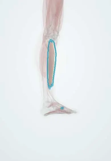
Tibialis anterior !
The tibialis anterior is the muscle along the front of the shin; it lifts the foot, and its weakness causes foot drop.
Official name: Tibialis anterior muscle
Opens in the Durgical app, no sign-up needed.
What it is
The muscle along the front of the shin.
What it does
Lifts the foot when walking.
Key facts
- Origin
- Lateral tibial condyle and upper half of lateral tibial shaft; interosseous membrane
- Insertion
- Medial and plantar surfaces of medial cuneiform; base of first metatarsal
- Action
- Dorsiflexes ankle; inverts foot
- Nerve
- Deep fibular nerve (L4–L5)
- Blood supply
- Anterior tibial artery
Relations
- Course
- Its tendon forms in the lower third of the leg, passes under the superior and inferior extensor retinacula in its own synovial sheath and crosses the front of the ankle to the medial side of the midfoot
- Medial
- The subcutaneous medial surface of the tibia (the shin)
- Lateral
- Extensor digitorum longus above and extensor hallucis longus below
- Deep
- Lateral surface of the tibia and the interosseous membrane; the anterior tibial vessels and deep fibular nerve lie on the membrane in the angle between it and its lateral neighbours
- Superficial
- The thick, unyielding deep fascia of the leg and the skin
How to find it
With the foot lifted and turned in, the tendon stands out as the largest cord in front of the ankle, just in front of the medial malleolus. The belly is felt just lateral to the sharp anterior border of the tibia.
Examination
- Heel walk: inability to keep the forefoot off the ground indicates dorsiflexor weakness (deep or common fibular nerve, L4–L5 roots)
- Resisted dorsiflexion and inversion: weakness with a normal tendon outline suggests a nerve or root cause; loss of the tendon outline in front of the ankle suggests a rupture
On imaging
Ultrasound shows its tendon as the largest in front of the ankle. A rupture, usually in older people near the insertion, shows a gap and a retracted stump that may be felt as a lump above the ankle; MRI confirms tears and tendinopathy. In foot drop, MRI can show denervation oedema of the anterior compartment. Compartment syndrome is diagnosed clinically and by pressure, not by imaging.
Clinical notes
- Anterior compartment syndrome: pain on passive plantarflexion of the ankle and toes, a tense shin, weak dorsiflexion and numbness in the first web space (deep fibular nerve)
- Foot drop from a common fibular nerve lesion at the fibular neck spares inversion (tibialis posterior, tibial nerve); an L5 root lesion weakens inversion too
- Spontaneous rupture in older adults is often painless: foot slap, a lump in front of the ankle and loss of the tendon outline
- A split transfer of its tendon to the lateral foot is used to balance a spastic varus foot
Anatomical variants
- The tendon may split into two bands to the medial cuneiform and the first metatarsal, or insert on only one of them; insertions further away are rare
Development
A dorsal (extensor) muscle mass derivative, supplied by the deep fibular nerve; the medial rotation of the lower limb in the seventh week brings this extensor group to the front of the leg.
Memory aid
In front of the ankle, medial to lateral: "Tom Has Very Nice Dancing Pigs" (Tibialis anterior, extensor Hallucis longus, Vessels, Nerve [deep fibular], extensor Digitorum longus, Peroneus tertius).
Related conditions
Test yourself
Think of the answer, then tap the question to see it.
What are the attachments and actions of the tibialis anterior?
From the lateral tibial condyle, upper lateral tibia and interosseous membrane to the medial cuneiform and first metatarsal base; it dorsiflexes the ankle and inverts the foot.
Which nerve supplies the tibialis anterior?
The deep fibular nerve (L4–L5); the tibialis anterior is the classic muscle for the L4 myotome.
What gait results from weakness of the tibialis anterior?
Foot drop, with a high-stepping gait and foot slap; heel walking is lost.
What are the signs of anterior compartment syndrome?
Pain on passive plantarflexion of the ankle and toes, a tense shin, weak dorsiflexion and numbness in the first web space (deep fibular nerve).
How does a common fibular nerve lesion differ from an L5 root lesion in foot drop?
A fibular neck lesion spares inversion (tibialis posterior, tibial nerve); an L5 root lesion weakens inversion too.
Educational information, not medical advice. For any symptom, see a health professional.
Its names
- Official Latin (TA2)
- musculus tibialis anterior TA2 2644
- Official Spanish (DeCS)
- Músculo tibial anterior
- English (TA2)
- Tibialis anterior muscle
Related
- Common fibular nerve
- Deep fibular nerve
- Anterior tibial artery
- Tibialis anterior tendon
- Medial cuneiform
- First metatarsal
- Tibia
Sources: Terminologia Anatomica (TA2); Anatomy, Bony Pelvis and Lower Limb: Tibialis Anterior Muscles; 8.5 Development of the Appendicular Skeleton (OpenStax (Rice University), 2022) · adapted, CC BY 4.0.
Spotted a mistake?We are always improving for the community. Tell us what is wrong and our team will review it.Report a mistake
Representative model, for educational purposes only.