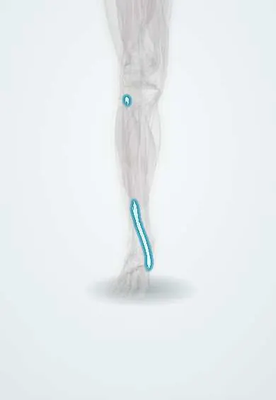
Tibialis anterior tendon !
The tibialis anterior tendon crosses the front of the ankle to the inner midfoot; it lifts the foot and is the most inner tendon there.
Official name: Tendon of tibialis anterior
Opens in the Durgical app, no sign-up needed.
What it is
The tendon of the shin muscle. It crosses the front of the ankle to the inner midfoot (the medial cuneiform and the first metatarsal).
What it does
Lifts the foot and helps hold up the inner arch.
Key facts
- Attaches
- Most medial tendon at the front of the ankle, under the extensor retinacula in its own compartment, to the medial and plantar surfaces of the medial cuneiform and the base of the first metatarsal
- Function
- Dorsiflexion and inversion; lowers the forefoot under control after heel strike; with the fibularis longus forms a stirrup under the midfoot
- Tight in
- Plantarflexion with eversion
Relations
- Course
- Down the lateral surface of the tibia, across the lower tibia to lie in front of the medial malleolus, then in its own sheath under the extensor retinacula to the inner midfoot
- Lateral
- Extensor hallucis longus tendon, then the anterior tibial artery and the deep fibular nerve
- Deep
- Lower tibia and the front of the ankle capsule
- Medial
- Great saphenous vein and saphenous nerve, passing in front of the medial malleolus
How to find it
With the foot lifted and turned in, it stands out as the thickest cord at the front of the ankle, on the inner side, running toward the inner midfoot.
Examination
- Resisted dorsiflexion and inversion: weakness with a missing tendon outline suggests rupture; walking on the heels is difficult
On imaging
Ultrasound and MRI show thickening near the insertion in tendinopathy, and a gap with a retracted stump, often near the extensor retinacula, in rupture.
Common problems
- Rarely, it tears in older adults, causing the foot to slap when walking.
Clinical notes
- In rupture the other dorsiflexors work and sensation is normal; in a deep fibular nerve palsy the toe extensors are weak too and the first web space may be numb
- After a clubfoot is corrected by casting, a foot that keeps turning in when walking (dynamic supination) is treated by moving the whole tendon to the lateral cuneiform
- The anterior approach to the ankle, used for ankle replacement and some plafond fractures, enters just lateral to this tendon, with the anterior tibial artery and deep fibular nerve kept lateral
Anatomical variants
Memory aid
Front of the ankle, medial to lateral: 'Tim Has A Nasty Dog' (Tibialis anterior, Hallucis longus, Artery, Nerve, Digitorum longus).
Test yourself
Think of the answer, then tap the question to see it.
What is the order of structures at the front of the ankle from medial to lateral?
Tibialis anterior, extensor hallucis longus, anterior tibial artery, deep fibular nerve, extensor digitorum longus ('Tim Has A Nasty Dog').
Where does the tibialis anterior tendon insert?
On the medial cuneiform and the base of the first metatarsal.
Why is tibialis anterior rupture in older adults often missed?
The toe extensors still lift the foot partly; clues are a lump above the ankle, lost tendon outline and a slapping gait.
How is tibialis anterior rupture told apart from deep fibular nerve palsy?
In rupture the toe extensors work and sensation is normal; in the nerve palsy the toe extensors are weak and the first web space may be numb.
Educational information, not medical advice. For any symptom, see a health professional.
Its names
- Official Latin (TA2)
- musculus tibialis anterior TA2 2644
- Official Spanish (DeCS)
- Tendón del tibial anterior DeCS D013710
- English (TA2)
- Tendon of tibialis anterior
Related
- Extensor retinacula
- Ankle tendon sheaths
- Ankle bursae
- Tibialis anterior
- Medial cuneiform
- First metatarsal
- Navicular
- Tibia
Sources: Terminologia Anatomica (TA2); DeCS; Anatomy, Bony Pelvis and Lower Limb: Tibialis Anterior Muscles.
Spotted a mistake?We are always improving for the community. Tell us what is wrong and our team will review it.Report a mistake
Representative model, for educational purposes only.