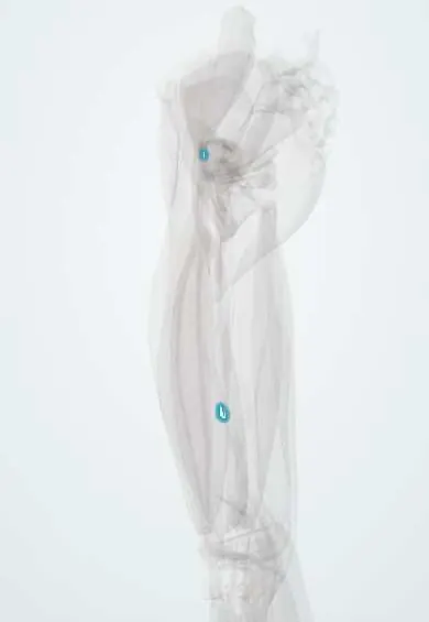
Adductor longus tendon !
The adductor longus tendon attaches this inner thigh muscle to the back of the femur, between the main thigh vessels.
Official name: Tendon of adductor longus
Opens in the Durgical app, no sign-up needed.
What it is
The lower end of the adductor longus on the back of the femur.
What it does
Pulls the leg inward.
Key facts
- Attaches
- Distal aponeurosis to the middle third of the linea aspera (medial lip), between the vastus medialis and the adductor magnus and brevis
- Function
- Carries the adduction pull to the femur; its other, proximal tendon on the pubic body is the usual site of adductor (groin) strain
- Tight in
- Hip abduction with extension
Relations
- In front
- Femoral artery and vein and, lower down, the adductor canal with the saphenous nerve
- Behind
- Deep femoral artery and vein, then the adductor brevis above and the adductor magnus below
- Lateral
- Vastus medialis, at the linea aspera
- Medial
- Gracilis
How to find it
The distal part is not palpable, deep in the inner thigh; it is found on MRI or in a medial approach between the femoral vessels in front and the adductor magnus behind. The proximal tendon, in contrast, is the prominent cord in the groin when the thigh is spread outward.
On imaging
The distal tendon is seen only on cross-sectional imaging: axial MRI or ultrasound of the mid-thigh show it as a dark band with the femoral vessels in front and the deep femoral vessels and adductor magnus behind. Distal tears are rare; in groin pain, imaging targets the other end, the proximal tendon at the pubis, where MRI shows oedema, a tear or an avulsion.
Clinical notes
- The deep femoral artery and its perforating arteries lie behind it near the linea aspera, where they can bleed with femoral shaft fractures, drilling or cerclage wiring
- In the adductor canal block for knee surgery, local anaesthetic is placed around the femoral artery deep to sartorius, where the canal is backed by the adductor longus and magnus; the saphenous nerve is the main target
Test yourself
Think of the answer, then tap the question to see it.
Which vessels does the distal adductor longus separate?
The femoral artery and vein in front from the deep femoral artery and vein behind.
Where on the femur does the adductor longus insert?
The middle third of the linea aspera (medial lip), between the vastus medialis and the adductor magnus and brevis.
Which end of the adductor longus is usually injured in a groin strain?
The proximal tendon and muscle–tendon junction near the pubis; distal injuries are rare.
Educational information, not medical advice. For any symptom, see a health professional.
Its names
- Official Latin (TA2)
- adductor longus TA2 2628
- Official Spanish (DeCS)
- Tendón del aductor largo DeCS D013710
- English (TA2)
- Tendon of adductor longus
- Also called
- adductor longus muscle
Related
Sources: Terminologia Anatomica (TA2); DeCS; Anatomy, Bony Pelvis and Lower Limb: Medial Thigh Muscles.
Spotted a mistake?We are always improving for the community. Tell us what is wrong and our team will review it.Report a mistake
Representative model, for educational purposes only.