Cardiovascular system · Lower limb
Femoral artery !
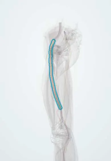
The femoral artery is the main artery of the thigh; its pulse is felt in the groin and it becomes the popliteal artery behind the knee.
Opens in the Durgical app, no sign-up needed.
What it is
The main artery of the thigh. It continues from the external iliac artery under the inguinal ligament, where its pulse can be felt in the groin, and runs down the front and inner thigh.
What it does
Brings blood to the leg. Its large branch, the deep femoral artery, feeds the thigh; the main trunk passes through the adductor canal and becomes the popliteal artery behind the knee.
Key facts
- Origin
- Continuation of external iliac artery behind inguinal ligament, at midinguinal point
- Branches
- Superficial epigastric, superficial circumflex iliac, external pudendals, deep femoral, descending genicular
- Supplies
- Whole lower limb; passes femoral triangle and adductor canal; becomes popliteal at adductor hiatus
Relations
- Course
- Enters the thigh behind the inguinal ligament in the lateral compartment of the femoral sheath, descends through the femoral triangle to its apex, runs in the adductor canal under the sartorius and leaves through the adductor hiatus to become the popliteal artery
- Medial
- The femoral vein in the middle compartment of the sheath and, beyond it, the femoral canal (fat, lymphatics, a lymph node), the route of a femoral hernia; lower down the vein moves behind the artery
- Lateral
- The femoral nerve, outside the sheath on the iliopsoas; in the adductor canal, the vastus medialis (the canal's lateral wall) and the nerve to vastus medialis
- Behind
- From above down: the psoas major tendon (separating it from the hip joint), the pectineus and the adductor longus; in the canal, the adductor longus and adductor magnus (its back wall)
- Superficial
- Skin, fascia lata and cribriform fascia in the triangle; in the canal, the sartorius over the vasto-adductor membrane (its roof), with the saphenous nerve crossing in front of it from lateral to medial
How to find it
With the person supine and the thigh slightly turned out, the pulse is felt just below the inguinal ligament at the mid-inguinal point, pressed against the femoral head. Lower down the sartorius covers it; its path follows the upper two-thirds of a line from the mid-inguinal point to the adductor tubercle.
Examination
- Radio-femoral delay: a femoral pulse that is weak and arrives after the radial pulse suggests coarctation of the aorta
- Ankle–brachial index (ankle systolic pressure divided by arm systolic pressure): below 0.9 indicates peripheral artery disease; a weak femoral pulse as well places the block at or above the groin
- Listening over the groin: a bruit points to turbulent flow through a narrowed iliac or common femoral artery
On imaging
Duplex ultrasound is first line: in the groin the artery lies lateral to the vein over the femoral head, with a triphasic waveform. Stenosis shows as high velocity and turbulence; occlusion as absent flow with collaterals. CT or MR angiography maps disease before surgery. Plain films only show wall calcification. Ultrasound also checks a puncture site for false aneurysm.
Common problems
- Narrowing or blockage from plaque (peripheral artery disease), causing leg pain when walking (claudication).
- It is the most common entry point for catheter procedures, such as heart catheterization; a bruise or false aneurysm can follow.
- Injury from deep cuts or fractures.
How it is diagnosed
Checking pulses; ankle-brachial index; duplex ultrasound; CT or catheter angiography.
Treatment
- Walking programs, quitting smoking and medicines for artery disease.
- Angioplasty and stenting, or surgical bypass, for severe narrowing.
- Repair of an injured or bulging artery.
Clinical notes
- Catheter access is over the femoral head, below the inguinal ligament, so the artery can be compressed against bone; a puncture above the ligament can bleed unseen into the retroperitoneum, one below the bifurcation risks a false aneurysm or arteriovenous fistula
- An embolus at the bifurcation gives a sudden painful, pale, pulseless leg; the clot can be removed through a groin incision with a balloon catheter (embolectomy)
- Femoro-popliteal bypass treats long blockages; the great saphenous vein is the preferred conduit
Anatomical variants
- The height of the deep femoral artery's origin varies and does not match the groin crease, so the crease is a poor guide for puncture
- Persistent sciatic artery (rare): the embryonic axial artery stays the main supply of the leg and the femoral artery is small
- Duplicated superficial femoral artery (rare)
Development
The first artery of the embryonic leg is the axial (sciatic) artery, which runs behind the thigh with the sciatic nerve. The femoral artery appears later in front, joins it above the knee and takes over; the axial vessel regresses, its lower part surviving in the fibular artery. When it persists, it is the persistent sciatic artery.
Memory aid
Groin from lateral to medial: NAVEL, Nerve, Artery, Vein, Empty space (femoral canal), Lymphatics.
Related conditions
- Familial dysbetalipoproteinemia (type III hyperlipoproteinemia)
- Peripheral artery disease
- Acute limb ischaemia
- Atherosclerosis and dyslipidaemia
Test yourself
Think of the answer, then tap the question to see it.
Where is the femoral pulse felt?
At the mid-inguinal point, halfway between the ASIS and the pubic symphysis, just below the inguinal ligament over the femoral head.
What is the order of structures in the groin from lateral to medial?
Nerve, artery, vein, empty space (femoral canal), lymphatics: NAVEL. The femoral vein lies medial to the femoral artery.
Where does the femoral artery become the popliteal artery?
At the adductor hiatus, after running through the femoral triangle and the adductor canal under the sartorius.
Why is femoral catheter access made over the femoral head, below the inguinal ligament?
The artery can be compressed against bone; a puncture above the ligament can bleed into the retroperitoneum, one below the bifurcation risks a false aneurysm.
Where is the commonest atherosclerotic blockage in the leg, and where does an embolus usually lodge?
Blockage: the superficial femoral artery near the adductor hiatus. Embolus: the femoral bifurcation.
Educational information, not medical advice. For any symptom, see a health professional.
Its names
- Official Latin (TA2)
- arteria femoralis TA2 4674
- Official Spanish (DeCS)
- Arteria femoral DeCS D005263
- English (TA2)
- Femoral artery
Related
- Popliteal artery
- Inguinal ligament
- External iliac artery
- Femoral vein
- Femoral nerve
- Deep femoral artery
- Rectus femoris
- Vastus medialis
Sources: Terminologia Anatomica (TA2); DeCS; Anatomy, Bony Pelvis and Lower Limb: Femoral Artery; Anatomy of the Human Body (20th ed.): Development of the Vascular System (Lea & Febiger (Gray's Anatomy, 1918), 1918) · adapted, Public domain; Anatomía humana (4.ª ed.), tomo 1, cap. 67: Arterias del miembro inferior.
Spotted a mistake?We are always improving for the community. Tell us what is wrong and our team will review it.Report a mistake
Representative model, for educational purposes only.