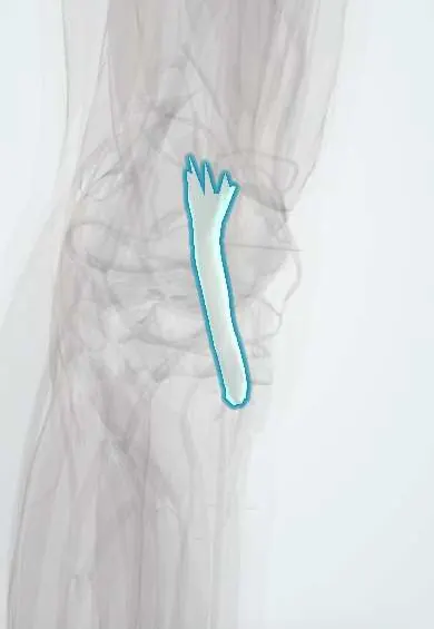
Vastus lateralis tendon !
The vastus lateralis tendon is the part of the quadriceps tendon that comes from the outer thigh muscle and attaches to the kneecap.
Official name: Tendon of vastus lateralis
Opens in the Durgical app, no sign-up needed.
What it is
The part of the quadriceps tendon that comes from the outer thigh muscle.
What it does
Transmits the force of the quadriceps to straighten the knee.
Key facts
- Attaches
- Middle layer of the quadriceps tendon to the lateral base and border of the patella; expansion to the lateral patellar retinaculum and tibial condyle
- Function
- Carries the vastus lateralis pull to the patella to extend the knee; pulls the patella laterally, balanced by the vastus medialis
- Tight in
- Knee flexion
Relations
- Superficial
- Rectus femoris tendon, the top layer, which it joins just above the patella
- Medial
- Vastus medialis tendon, with which it shares the middle layer
- Deep
- Vastus intermedius tendon, the deep layer, over the suprapatellar bursa
- Lateral
- Iliotibial band (IT band) and the lateral retinaculum, which its expansion strengthens
How to find it
With the knee straight and the thigh tightened, it is felt as the firm outer margin of the quadriceps tendon just above the upper outer corner of the patella.
On imaging
On sagittal and axial MRI the quadriceps tendon usually shows separate layers divided by thin fat lines (often three, sometimes two or four); a partial tear may involve one layer only, most often the superficial rectus femoris fibres.
Common problems
- Tendinitis from repeated jumping or kneeling.
- Rupture, usually in older adults; the leg cannot be straightened.
How it is diagnosed
Physical exam; ultrasound or MRI.
Treatment
- Tendinitis: rest, physical therapy.
- Complete tear: surgical repair, usually soon after the injury.
- Part of this tendon can also be used as an ACL graft.
Clinical notes
- Lateral retinacular release, once common for patellar maltracking, cuts through its expansion; an excessive release can leave the patella unstable medially
- A complete quadriceps tendon rupture often extends into the medial and lateral expansions; when these retinacular fibres are also torn, the person cannot lift the straight leg against gravity
Anatomical variants
- The lowest fibres often form a separate oblique part (vastus lateralis obliquus) reaching the lateral retinaculum and patella; one review found two insertional parts in about 60%
Test yourself
Think of the answer, then tap the question to see it.
Which layer of the quadriceps tendon does the vastus lateralis tendon form?
The lateral half of the middle layer, with the vastus medialis; rectus femoris is on top and vastus intermedius deepest.
Why does the vastus lateralis tendon favour lateral patellar instability?
It pulls the patella upward and outward, balanced by the vastus medialis.
Which retinaculum does the vastus lateralis tendon reinforce?
The lateral patellar retinaculum; an excessive lateral release through it can leave the patella unstable medially.
Educational information, not medical advice. For any symptom, see a health professional.
Its names
- Official Latin (TA2)
- musculus vastus lateralis TA2 2618
- Official Spanish (DeCS)
- Tendón del vasto lateral DeCS D013710
- English (TA2)
- Tendon of vastus lateralis
- Also called
- musculus vastus fibularis
Related
Sources: Terminologia Anatomica (TA2); DeCS; Anatomy, Bony Pelvis and Lower Limb: Vastus Lateralis Muscle; Anatomy, Bony Pelvis and Lower Limb: Thigh Quadriceps Muscle.
Spotted a mistake?We are always improving for the community. Tell us what is wrong and our team will review it.Report a mistake
Representative model, for educational purposes only.