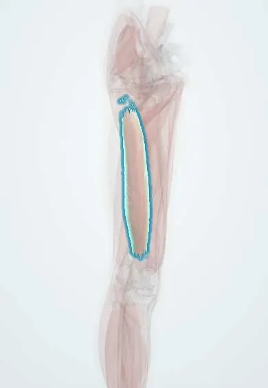
Vastus intermedius !
The vastus intermedius is the deepest quadriceps muscle, lying directly on the femur under the others; it straightens the knee.
Official name: Vastus intermedius muscle
Opens in the Durgical app, no sign-up needed.
What it is
The deepest quadriceps muscle, lying on the front of the femur.
What it does
Straightens the knee.
Key facts
- Origin
- Anterior and lateral surfaces of upper two-thirds of femoral shaft
- Insertion
- Deep surface of quadriceps tendon; base of patella; tibial tuberosity via patellar ligament
- Action
- Extends knee
- Nerve
- Femoral nerve (L2–L4)
- Blood supply
- Lateral circumflex femoral artery; deep femoral artery
Relations
- Superficial
- Rectus femoris
- Deep
- Anterior and lateral surfaces of the femoral shaft
- Medial
- Vastus medialis, partly blended with it
- Lateral
- Vastus lateralis, partly blended with it
- Below
- Articularis genus and the suprapatellar bursa, between the quadriceps tendon and the front of the femur
How to find it
It cannot be felt on its own. On transverse ultrasound of the mid-thigh it is the layer lying directly on the bright femoral cortex, under the rectus femoris; both are measured to follow quadriceps wasting.
Examination
- Patellar (knee-jerk) reflex: a tap on the patellar ligament contracts all four quadriceps heads; a reduced or absent reflex points to the femoral nerve or the L3–L4 roots (mainly L4)
On imaging
On transverse ultrasound of the mid-thigh it is the deepest layer, lying on the bright femoral cortex under the rectus femoris; the two are measured together to track quadriceps wasting. After a direct blow, ultrasound or MRI show a haematoma inside it. If a firm lump persists, radiographs after a few weeks show the faint rim of calcification of myositis ossificans, which CT confirms.
Common problems
- Strains (pulled muscle) from sprinting or kicking; bruising from a direct blow.
- Weakness and shrinking after knee injury or surgery, which rehabilitation works to rebuild.
How it is diagnosed
Physical exam; ultrasound or MRI for significant injuries.
Treatment
- Rest, ice, then progressive strengthening with a physical therapist.
Clinical notes
- After a femoral shaft fracture or thigh surgery it can scar to the bone and limit knee bending; a surgical release of the quadriceps (quadricepsplasty) may then be needed
- The severity of a thigh contusion is commonly graded by how much knee flexion is still possible in the first days
Anatomical variants
- Tensor of the vastus intermedius: an extra muscle belly between the vastus lateralis and the vastus intermedius, described in several forms
Development
From the dorsal muscle mass, like the other quadriceps heads; its deep slips form the articularis genus, which lifts the suprapatellar bursa, itself an upward pouch of the knee joint cavity.
Related conditions
Test yourself
Think of the answer, then tap the question to see it.
Why is the vastus intermedius typically involved in a thigh contusion and later myositis ossificans?
It is the deepest quadriceps head, lying directly on the femur, so a direct blow crushes it against the bone.
Which slips from the deep surface of the vastus intermedius lift the suprapatellar bursa as the knee straightens?
The articularis genus, often described as a separate muscle.
After a femoral shaft fracture, how can the vastus intermedius limit knee movement?
It can scar to the bone and limit knee bending; a surgical release of the quadriceps (quadricepsplasty) may then be needed.
Educational information, not medical advice. For any symptom, see a health professional.
Its names
- Official Latin (TA2)
- musculus vastus intermedius TA2 2619
- Official Spanish (DeCS)
- Músculo vasto intermedio DeCS D052097
- English (TA2)
- Vastus intermedius muscle
Related
- Rectus femoris
- Vastus medialis
- Vastus lateralis
- Femoral nerve
- Femoral artery
- Vastus intermedius tendon
- Femur
Sources: Terminologia Anatomica (TA2); DeCS; Anatomy, Bony Pelvis and Lower Limb: Thigh Quadriceps Muscle.
Spotted a mistake?We are always improving for the community. Tell us what is wrong and our team will review it.Report a mistake
Representative model, for educational purposes only.