Conditions · Ophthalmology and ENT
Endophthalmitis !
A severe infection inside the eye, usually after eye surgery, an injection or a penetrating injury. It is an emergency that can cause blindness within hours.
Updated 2026-11-01
Structures involved
Tap one to see what it is and open it in 3D.
Why it happens
- Patterns of acute inflammation: serous, fibrinous, purulent, abscess, ulcer
- The eye wall: cornea, sclera, uvea and lens
What it is
Inflammation, almost always infectious, of the internal cavities of the eye: the vitreous and the aqueous humour, often spreading to the retina and uvea. Exogenous endophthalmitis follows entry of organisms from outside (surgery, intravitreal injection, trauma, corneal ulcer); endogenous endophthalmitis is seeded through the bloodstream from an infection elsewhere.
Who gets it
Rare but feared. Acute postoperative endophthalmitis follows roughly one in a thousand cataract operations or fewer, and post-injection cases are rarer still; because cataract surgery and intravitreal injections are very common, these are the main sources. Open-globe injuries, frequent in agricultural and work-related trauma in Latin America, carry a much higher risk, especially with retained foreign bodies or soil contamination. Endogenous cases are a small minority and occur in seriously ill or immunocompromised patients.
Causes
- Acute postoperative (days after surgery): mainly coagulase-negative staphylococci, also Staphylococcus aureus and streptococci
- Chronic postoperative (weeks to months): low-virulence organisms such as Cutibacterium (Propionibacterium) acnes, or fungi
- Post-traumatic: Bacillus cereus (rapid and destructive), staphylococci and fungi after injuries with soil or plant matter
- Bleb-related after glaucoma surgery: streptococci and Haemophilus
- Endogenous: Candida and other fungi, or bacteria such as Klebsiella, S. aureus and streptococci from a distant focus
Risk factors
- Recent intraocular surgery, especially with a posterior capsule rupture or a leaking wound
- Intravitreal injection
- Penetrating eye injury, particularly with a retained intraocular foreign body or rural setting
- Glaucoma filtering bleb
- Endogenous: diabetes, intravenous drug use, central venous catheters, total parenteral nutrition, immunosuppression, endocarditis and liver abscess
Pathophysiology
Organisms reach the normally sterile vitreous, a gel that offers little defence and is easily colonised. Bacterial toxins and the host's neutrophilic response produce pus in the anterior chamber (hypopyon) and vitreous, and inflammatory mediators damage the retina within hours. The retina does not regenerate, so delay translates directly into permanent vision loss; severe cases spread to all coats of the eye (panophthalmitis) and the orbit.
Classification
- Exogenous: acute postoperative, chronic (delayed) postoperative, post-injection, bleb-related, post-traumatic, keratitis-related
- Endogenous (haematogenous): bacterial or fungal
- Panophthalmitis: infection extending through the sclera into orbital tissue
Symptoms and signs
- Decreased vision, often marked, in an eye operated or injected days earlier
- Eye pain that worsens instead of improving after surgery
- Red eye with eyelid swelling and conjunctival oedema
- Hypopyon (layer of pus in the anterior chamber) and cells in the anterior chamber and vitreous
- Loss of the red reflex and a hazy view of the fundus
- Endogenous: often a fever and a systemic infection, sometimes in both eyes
Diagnosis
- Clinical
- Mainly clinical: inflammation out of proportion to the expected postoperative course, with hypopyon, vitritis and reduced red reflex. Visual acuity at presentation is recorded because it guides treatment and prognosis.
- Tests
- Samples of vitreous (preferred) and aqueous humour for Gram stain, culture and, where available, PCR before or at the moment of intravitreal treatment. In endogenous cases, blood cultures, echocardiography and a search for the source.
- Imaging
- B-scan ultrasound when the view of the retina is blocked, to show vitreous opacities, retinal detachment or a foreign body. CT of the orbit after trauma to locate a metallic foreign body (MRI is avoided if metal is suspected).
Differential diagnosis
- Toxic anterior segment syndrome (sterile inflammation within 24 hours of surgery, no vitreous involvement)
- Retained lens fragments with postoperative uveitis
- Non-infectious uveitis
- Infectious keratitis with a sterile hypopyon
- Orbital cellulitis (proptosis and limited eye movements rather than intraocular pus)
Goal of treatment
Sterilise the inside of the eye as quickly as possible to save vision and the eye, and treat any systemic source.
Treatment
- Acute
- Emergency referral to ophthalmology; intraocular samples are taken and broad-spectrum intravitreal antibiotics covering gram-positive and gram-negative bacteria are injected the same day.
- Surgical
- Pars plana vitrectomy removes infected vitreous and is favoured when vision is light perception only, in severe or post-traumatic cases, or when the eye does not improve.
- Medical
- Intravitreal antifungals plus systemic antifungals when fungi are suspected; systemic antibiotics as an adjunct in post-traumatic and endogenous cases; steroids are debated and avoided when fungal infection is possible.
- Medical
- Endogenous disease: treat the primary infection (endocarditis, abscess, candidaemia) and remove infected catheters.
- Follow-up
- Daily review of pain, acuity and hypopyon in the first days, with repeat injection if there is no improvement in about 48 hours; nursing care includes eye protection, pain control and teaching the patient to report worsening at once.
Complications
- Permanent vision loss or blindness
- Retinal detachment
- Panophthalmitis and orbital spread
- Phthisis bulbi (shrunken, non-functional eye) or need for evisceration
- Spread of infection in endogenous disease
Prognosis
Depends on the organism and on vision at presentation. Low-virulence staphylococcal infections treated promptly often recover useful vision; streptococci, Bacillus and late treatment carry a poor outlook, and many endogenous cases end with severe loss.
Prevention
- Antiseptic preparation of the eye surface with povidone-iodine before surgery and injections
- Intracameral antibiotic at the end of cataract surgery, as used in many centres
- Watertight wound closure and prompt repair of open-globe injuries with early antibiotics
- Protective eyewear for agricultural and industrial work
- Routine eye examination in candidaemia
Test yourself
Think of the answer, then tap the question to see it.
A patient has pain, decreased vision and hypopyon 4 days after cataract surgery. Most likely diagnosis?
Acute postoperative endophthalmitis.
Most common organisms in acute postoperative endophthalmitis?
Coagulase-negative staphylococci.
What sample is preferred to identify the organism?
Vitreous, for Gram stain, culture and PCR.
Which organism causes a fulminant endophthalmitis after penetrating trauma with soil contamination?
Bacillus cereus.
In a patient with candidaemia, why is a fundus examination done?
To detect endogenous Candida endophthalmitis, which changes treatment.
Related conditions
- Cataract
- Keratitis and corneal ulcer
- Ocular trauma: hyphema and open globe injury
- Uveitis
- Candidiasis: oral, oesophageal and invasive
- Infective endocarditis
- Orbital cellulitis
- Sepsis and septic shock
- Conjunctivitis
- Diabetic retinopathy
Educational information, not medical advice. For any symptom, see a health professional.
Sources: What is Endophthalmitis? (American Academy of Ophthalmology, 2026); Endophthalmitis (EyeWiki, American Academy of Ophthalmology, 2026).
Spotted a mistake?We are always improving for the community. Tell us what is wrong and our team will review it.Report a mistake
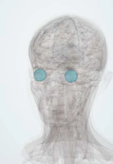
Representative model, for educational purposes only.