Conditions · Metabolism (biochemistry)
Amyloidosis !
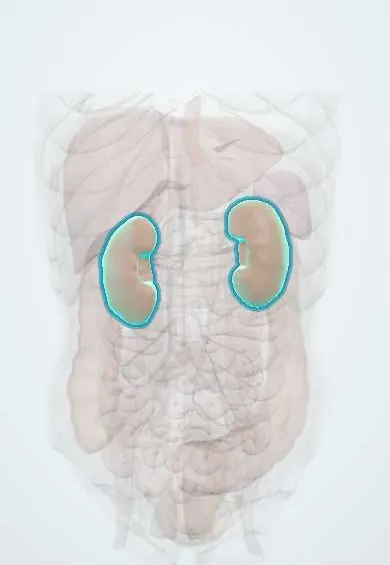
Misfolded proteins form insoluble fibres (amyloid) that build up in the heart, kidney, nerves and other organs and make them fail.
Updated 2026-11-01
Structures involved
Tap one to see what it is and open it in 3D.
In biochemistry
- Molecular chaperones and heat shock proteins
- Proteasome (26S, β5 subunit)
- Protein structure and folding
- Plasma proteins and electrophoresis
- Immunoglobulins (antibodies)
- Transthyretin (prealbumin)
Why it happens
- Special stains in diagnosis: Congo red, Prussian blue, PAS, Ziehl–Neelsen, Gram
- Systemic effects of inflammation: fever, acute-phase proteins, leukocytosis, sepsis
- Endoplasmic reticulum stress and the unfolded protein response
- Chronic inflammation: causes, cells and features
What it is
A group of diseases in which a misfolded plasma protein is laid down outside cells as cross-β-sheet fibrils; each type is named after its protein (AL, AA, ATTR…).
Who gets it
Uncommon. AL is the commonest systemic type in high-income countries; wild-type ATTR is increasingly recognised in older men with heart failure. No Colombia-specific figures were found.
Causes
- AL: immunoglobulin light chains from a plasma-cell clone (sometimes with myeloma)
- AA: serum amyloid A protein in chronic inflammation (rheumatoid arthritis, tuberculosis, familial Mediterranean fever)
- ATTR: variant (hereditary) or normal (older adults) transthyretin
- Others: β2-microglobulin (long-term dialysis), β-amyloid (Alzheimer, localised), amylin (islets in type 2 diabetes)
Risk factors
- Monoclonal gammopathy (MGUS, myeloma)
- Chronic inflammatory diseases
- TTR variants and family history
- Older age and male sex (wild-type ATTR)
- Many years of haemodialysis
Pathophysiology
The precursor protein loses its native shape (from an unstable variant, overproduction or age), exposes segments that stack into β-sheets and forms fibrils that resist breakdown. The fibrils settle in the extracellular matrix with serum amyloid P component and glycosaminoglycans, thicken walls and push cells apart: the myocardium stiffens (restrictive cardiomyopathy), the glomerulus leaks protein (nephrotic syndrome) and nerves are compressed or degenerate. Pre-fibrillar oligomers are toxic too.
Classification
- AL (light chains)
- AA (serum amyloid A)
- Hereditary ATTR and wild-type ATTR
- Localised (cerebral β-amyloid, amylin, calcitonin in medullary thyroid carcinoma)
Symptoms and signs
- Breathlessness, oedema and fatigue from heart failure with thick walls
- Foamy urine and oedema from nephrotic syndrome
- Tingling and pain in feet and hands, bilateral carpal tunnel syndrome
- Dizziness on standing, diarrhoea or constipation (autonomic neuropathy)
- Macroglossia and bruising around the eyes (typical of AL)
- Weight loss and hepatomegaly
Diagnosis
- Clinical
- Suspect it with heart failure plus thick walls and low voltage, unexplained nephrotic syndrome, neuropathy with bilateral carpal tunnel, or macroglossia.
- Tests
- Biopsy (abdominal fat, salivary gland or affected organ) with Congo red: apple-green birefringence under polarised light; typing by immunohistochemistry or mass spectrometry; serum and urine electrophoresis and immunofixation and free light chains; proteinuria, troponin and NT-proBNP; electrocardiogram (low voltage); TTR genetic testing.
- Imaging
- Echocardiography (thick walls, restrictive pattern, strain with apical sparing); cardiac MRI with diffuse late enhancement; technetium pyrophosphate bone scintigraphy for ATTR.
Treatment
- Medical
- AL: treatment aimed at the plasma-cell clone (proteasome inhibitors, anti-CD38 antibodies, corticosteroids)
- Procedure
- AL: autologous haematopoietic stem cell transplant in selected patients
- Medical
- ATTR: transthyretin stabilisers (tafamidis, acoramidis) or silencers of its synthesis (patisiran, vutrisiran)
- Medical
- AA: control the underlying inflammatory disease (colchicine in familial Mediterranean fever, biologics in rheumatoid arthritis)
- Conservative
- Management of heart and kidney failure with diuretics and fluid control; some cardiac drugs are poorly tolerated
- Surgical
- Heart, kidney or liver transplant in selected cases
Complications
- Heart failure and sudden death
- End-stage kidney disease
- Disabling neuropathy
- Orthostatic hypotension
Prognosis
Depends on the type and on cardiac damage at diagnosis; advanced heart involvement worsens the outlook greatly. Targeted treatments have improved survival.
Test yourself
Think of the answer, then tap the question to see it.
Which stain confirms amyloid in a biopsy?
Congo red, which shows apple-green birefringence under polarised light because of the cross-β-sheet structure.
Which protein does AL amyloid come from, and what is it linked to?
Immunoglobulin light chains from a plasma-cell clone; it is linked to gammopathies and myeloma.
Which amyloidosis develops in chronic inflammation?
AA, from serum amyloid A, an acute-phase reactant the liver overproduces.
Which imaging test points to transthyretin cardiac amyloidosis?
Technetium pyrophosphate scintigraphy, which shows intense uptake in ATTR myocardium.
Related conditions
- Multiple myeloma
- Prion diseases (Creutzfeldt–Jakob)
- Heart failure
- Carpal tunnel syndrome
- Alzheimer disease
Educational information, not medical advice. For any symptom, see a health professional.
Sources: Amyloidosis: MedlinePlus (MedlinePlus, US National Library of Medicine, 2026); Primary amyloidosis: MedlinePlus Medical Encyclopedia (MedlinePlus, US National Library of Medicine, 2026); Transthyretin amyloidosis: MedlinePlus Genetics (MedlinePlus, US National Library of Medicine, 2026); TTR gene: MedlinePlus Genetics (MedlinePlus, US National Library of Medicine, 2026).
Spotted a mistake?We are always improving for the community. Tell us what is wrong and our team will review it.Report a mistake
Representative model, for educational purposes only.