
Aniridia and anterior segment dysgenesis
Inborn faults in how the front of the eye forms: missing iris (aniridia), Axenfeld-Rieger syndrome or Peters anomaly. All carry a high glaucoma risk.
What it is
Congenital neural crest disorders of the cornea, iris and angle. Aniridia (PAX6) affects the whole eye: iris and foveal hypoplasia with nystagmus. Axenfeld-Rieger syndrome (PITX2, FOXC1) adds iris strands to the angle and dental and facial anomalies. Peters anomaly is a central corneal opacity with iris or lens adhesions.
Who gets it
Each is rare (aniridia about 1 in 40 000 to 100 000 births); about two thirds of aniridia is inherited as autosomal dominant, and the rest is sporadic, sometimes as WAGR syndrome.
Causes
- Aniridia: PAX6 mutation or deletion on chromosome 11p13
- WAGR syndrome: deletion of 11p13 including PAX6 and WT1 (Wilms tumour, aniridia, genitourinary anomalies, intellectual disability)
- Axenfeld-Rieger syndrome: PITX2 or FOXC1 mutations, autosomal dominant
- Peters anomaly: mostly sporadic; PAX6, PITX2, FOXC1, CYP1B1 and other genes; Peters-plus syndrome (B3GLCT)
Risk factors
- Affected parent (autosomal dominant forms)
- Sporadic aniridia, which carries the WAGR risk
Pathophysiology
Faulty migration or differentiation of neural crest cells leaves the iris, angle and corneal endothelium incomplete. Abnormal drainage structures cause glaucoma, and in aniridia limbal stem cell deficiency gives progressive keratopathy.
Classification
- Aniridia (isolated or WAGR)
- Axenfeld anomaly: posterior embryotoxon (prominent, displaced Schwalbe line) with iris strands to it
- Rieger anomaly: Axenfeld findings plus iris hypoplasia, corectopia and holes
- Rieger syndrome: ocular findings plus dental anomalies, maxillary hypoplasia and redundant periumbilical skin
- Peters anomaly type 1 (iridocorneal adhesions) and type 2 (lens adherent to the cornea, often bilateral and severe)
Symptoms and signs
- Absent or irregular iris with photophobia
- Nystagmus and poor vision from infancy (aniridia)
- White cornea at birth (Peters anomaly)
- Watering, light sensitivity and a large cornea if congenital glaucoma develops
Diagnosis
- Clinical
- Examination under anaesthesia if needed: iris, cornea, gonioscopy, pressure, lens and fundus (foveal hypoplasia); examination of the parents; dental and systemic examination.
- Tests
- Genetic testing (PAX6, chromosomal microarray for 11p13 deletion including WT1, PITX2, FOXC1); pressure and corneal diameter measurement.
- Imaging
- Renal ultrasound every few months until about age 8 in WAGR risk; anterior segment OCT or ultrasound biomicroscopy for the angle and cornea; OCT for foveal hypoplasia.
Differential diagnosis
- Iris coloboma
- Traumatic or surgical iris loss
- Iridocorneal endothelial syndrome (acquired, unilateral)
- Congenital glaucoma, birth trauma or infection as other causes of a cloudy infant cornea
Inborn anterior segment disorders compared
| Feature | Aniridia | Axenfeld-Rieger | Peters anomaly | Primary congenital glaucoma |
|---|---|---|---|---|
| Main genes | PAX6 (11p13) | PITX2, FOXC1 | Mostly sporadic; PAX6, PITX2, FOXC1, CYP1B1 | CYP1B1 in many familial cases |
| Key eye sign | Iris almost absent, only a stump | Posterior embryotoxon with iris strands, corectopia, iris holes | Central corneal opacity with iris or lens adhesion | Large, cloudy cornea with a normal iris |
| Fovea and nystagmus | Foveal hypoplasia and nystagmus | Fovea normal | Fovea normal; amblyopia from the opacity | Fovea normal |
| Body beyond the eye | Wilms tumour and genitourinary anomalies in WAGR | Teeth, maxilla, redundant periumbilical skin | Peters-plus syndrome in some | Usually none |
| Glaucoma | Up to half | Up to half | Common | The defining feature |
Goal of treatment
Preserve vision by controlling glaucoma, protecting the cornea and treating amblyopia, and detect systemic associations early.
Treatment
- Medical
- Pressure-lowering drops; lubricants for aniridic keratopathy.
- Surgical
- Angle surgery, trabeculectomy or drainage devices for glaucoma; corneal transplant or limbal stem cell grafting in selected cases; early keratoplasty or optical iridectomy in bilateral Peters anomaly.
- Rehabilitation
- Tinted or iris-printed contact lenses for glare, spectacles, amblyopia therapy and low-vision support.
- Follow-up
- Lifelong pressure checks; regular renal ultrasound for children with WT1 deletion.
Complications
- Glaucoma (up to half of patients with aniridia or Axenfeld-Rieger)
- Aniridia-associated keratopathy
- Cataract
- Amblyopia
- Wilms tumour in WAGR syndrome
Common traps
- Aniridia is only a missing iris: PAX6 acts on the whole eye, so foveal hypoplasia, nystagmus, keratopathy and cataract decide the vision, not the iris gap
- Every child with aniridia carries the same Wilms tumour risk: the risk lies in sporadic cases whose 11p13 deletion includes WT1, which is why microarray guides the renal ultrasound schedule
- A prominent Schwalbe line alone means Axenfeld-Rieger syndrome: an isolated posterior embryotoxon is seen in some normal eyes; the anomaly needs iris strands to it or other angle and iris defects
- A white cornea in a newborn is Peters anomaly until proven otherwise: congenital glaucoma, birth trauma and infection also cloud the cornea, and glaucoma needs pressure and corneal diameter measured at once
- Glaucoma in these children is a childhood-only problem: it can appear at any age, so pressure checks are lifelong
Prognosis
Vision is usually limited in aniridia because of foveal hypoplasia; outcome depends on glaucoma control and corneal health.
Test yourself
Think of the answer, then tap the question to see it.
An infant has sporadic aniridia and no family history. Which test decides how closely the kidneys are watched, and why?
Chromosomal microarray for an 11p13 deletion. If WT1 is deleted, Wilms tumour risk is high and renal ultrasound every few months until about age 8 is needed.
Why does a child with aniridia see poorly even with a contact lens that mimics an iris?
PAX6 loss also leaves the fovea underdeveloped, causing nystagmus and low acuity; the iris lens only cuts glare.
A child has posterior embryotoxon with iris strands, missing teeth and redundant periumbilical skin. Which diagnosis, and what is the main eye risk?
Axenfeld-Rieger syndrome (PITX2 or FOXC1); glaucoma develops in up to half, so pressure is checked for life.
Why does aniridic keratopathy worsen over the years?
Limbal stem cells are deficient, so the corneal surface cannot renew and conjunctiva grows over it; lubricants help and limbal stem cell grafts are used in selected cases.
A newborn has dense central white corneas with iris adhesions in both eyes. Why is early surgery considered?
Bilateral Peters anomaly blocks the visual axis during the critical period, so early keratoplasty or optical iridectomy aims to prevent dense amblyopia.
Connections
Educational information, not medical advice. For any symptom, see a health professional.
Sources: Aniridia (StatPearls Publishing (NCBI Bookshelf), 2023); Axenfeld-Rieger Syndrome (StatPearls Publishing (NCBI Bookshelf), 2024); Peters Anomaly (StatPearls Publishing (NCBI Bookshelf), 2023).
Spotted a mistake?We are always improving for the community. Tell us what is wrong and our team will review it.Report a mistake
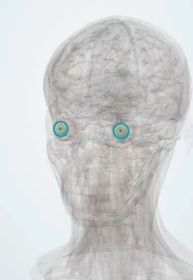
Representative model, for educational purposes only.