
Nystagmus
Involuntary rhythmic movements of the eyes. In infants it often reveals poor vision; new nystagmus in adults points to the inner ear or the brain.
What it is
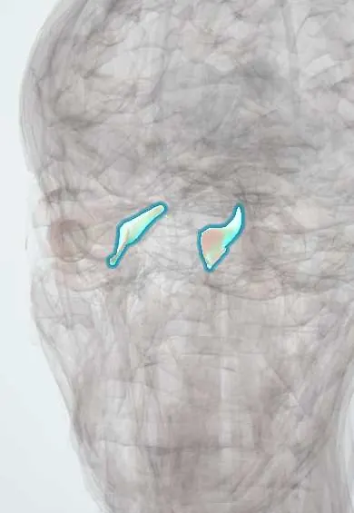
Involuntary, rhythmic oscillation of the eyes. It can be physiological (a few beats at the extremes of gaze), present from infancy, or acquired from disease of the inner ear, brainstem or cerebellum, drugs or alcohol. Its timing, direction and response to fixation help locate the cause.
Who gets it
Infantile nystagmus appears in the first months of life; acquired nystagmus is common in vestibular disorders and neurological disease.
Causes
- Infantile: idiopathic, or from poor vision (cataract, albinism, optic nerve or retinal disease)
- Peripheral vestibular disease (vestibular neuritis, benign positional vertigo)
- Central causes: stroke, multiple sclerosis, tumours, cerebellar degeneration
- Drugs (anticonvulsants, sedatives) and alcohol
- Thiamine deficiency (Wernicke encephalopathy)
Risk factors
- Visual impairment in infancy
- Neurological disease
- Anticonvulsant drugs and alcohol
Pathophysiology
Steady gaze depends on balanced input from both inner ears, the brainstem gaze-holding networks, the cerebellum and clear vision. Asymmetric vestibular input or failure of these networks lets the eyes drift, followed by corrective fast phases (jerk nystagmus) or a smooth to-and-fro motion (pendular nystagmus).
Classification
- Infantile or acquired
- Jerk or pendular
- Peripheral (vestibular) or central
Symptoms and signs
- Visible eye movements
- Oscillating vision (oscillopsia) in acquired forms
- Vertigo and nausea in vestibular causes
- Reduced vision
- Head turn to a position where the nystagmus is least
Diagnosis
- Clinical
- Describe direction, waveform, effect of gaze and of fixation; bedside head impulse and positional tests in acute vertigo; full eye and neurological exam.
- Tests
- Drug levels when relevant; electrophysiology in infants with suspected retinal disease.
- Imaging
- MRI of the brain for acquired central nystagmus or atypical features.
Differential diagnosis
- Physiological end-gaze nystagmus
- Saccadic intrusions (opsoclonus, square-wave jerks)
- Voluntary nystagmus
- Ocular flutter
Goal of treatment
Find and treat the cause, and reduce symptoms when no cause can be treated.
Treatment
- Acute
- Urgent stroke workup for central acute vestibular syndrome; thiamine at once when Wernicke encephalopathy is possible.
- Medical
- Adjusting or stopping causative drugs.
- Conservative
- Optical correction and low vision support in infantile nystagmus.
- Surgical
- Eye muscle surgery to reduce a head turn in selected infantile cases.
Complications
- Reduced visual acuity
- Oscillopsia
- Abnormal head posture
Prognosis
Depends on the cause; infantile nystagmus is lifelong but often improves with age, and peripheral vestibular nystagmus settles within days to weeks.
Test yourself
Think of the answer, then tap the question to see it.
Which features of nystagmus suggest a central cause?
Vertical or direction-changing nystagmus, or nystagmus that is not suppressed by fixation.
What must be looked for in an infant with nystagmus?
Causes of poor vision such as cataract, albinism, and retinal or optic nerve disease.
Which deficiency causes nystagmus with confusion and ataxia?
Thiamine deficiency (Wernicke encephalopathy).
In ophthalmology
Connections
Educational information, not medical advice. For any symptom, see a health professional.
Sources: Nystagmus Types (StatPearls Publishing (NCBI Bookshelf), 2025).
Spotted a mistake?We are always improving for the community. Tell us what is wrong and our team will review it.Report a mistake
Representative model, for educational purposes only.