Conditions · Pediatrics and neonatology
Wilms tumour (nephroblastoma) !
Wilms tumour is the most common kidney cancer in young children, usually found as a painless belly mass. Most children are cured with treatment.
Updated 2026-11-01
Structures involved
Tap one to see what it is and open it in 3D.
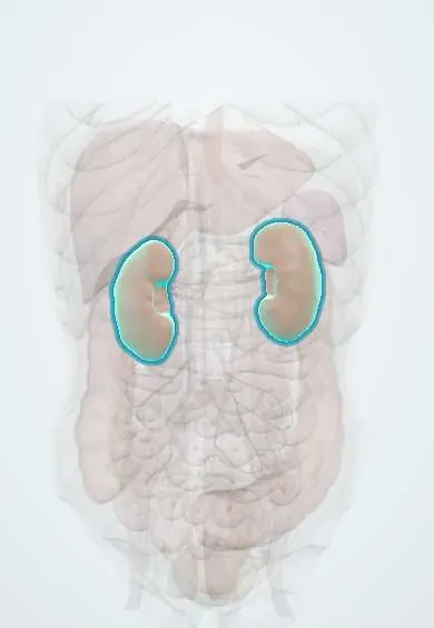
- Kidneys
- Renal pelvis
- Left renal vein
- Right renal vein
- Inferior vena cava
- Abdominal aorta
- Left adrenal gland
- Iris
In embryology
In the clinical exam
Why it happens
- Tumour suppressor genes: RB, TP53, APC, BRCA and the two-hit hypothesis
- Tumours of infancy and childhood
- Genomic imprinting: Prader–Willi and Angelman
- Naming tumours: -oma, carcinoma, sarcoma, and the misleading names
What it is
A malignant embryonal tumour of the kidney arising from persistent primitive renal tissue (nephrogenic rests). It is the most common renal cancer of childhood, typically in children aged 2–5 years.
Who gets it
Accounts for about 5–6% of childhood cancers, with most cases before 5 years of age; about 5–10% are bilateral. In Latin America, children are more often diagnosed at advanced stages and access to timely treatment affects survival; Colombia includes childhood cancer in its national surveillance and care programmes.
Causes
- Mostly sporadic
- Mutations in WT1 (11p13) or abnormalities at 11p15 (WT2) in syndromic cases
Risk factors
- WAGR syndrome (Wilms tumour, aniridia, genitourinary anomalies, intellectual disability)
- Denys-Drash and Frasier syndromes (WT1)
- Beckwith-Wiedemann syndrome and isolated hemihypertrophy
- Genitourinary anomalies such as cryptorchidism and hypospadias
- Family history of Wilms tumour
Pathophysiology
Embryonic kidney cells fail to mature and keep proliferating. The tumour typically has three components (blastemal, stromal and epithelial) and grows as a well-circumscribed mass within the kidney. It can extend into the renal vein and inferior vena cava, and spreads mainly to the lungs. Anaplastic histology predicts a worse response to treatment.
Classification
- Favourable histology (most cases) versus anaplastic histology
- Staging I–V (stage V: bilateral disease), used together with histology to guide therapy
Symptoms and signs
- Painless, smooth abdominal or flank mass, often noticed by a parent while bathing or dressing the child
- Abdominal pain or swelling
- Haematuria
- Hypertension (renin secretion)
- Fever, poor appetite or weight loss
- Aniridia, hemihypertrophy or genital anomalies as clues to a syndrome
Diagnosis
- Clinical
- A unilateral flank mass that does not usually cross the midline in a well-looking young child. Palpation should be gentle and limited, to avoid rupturing the tumour.
- Tests
- Complete blood count, kidney function, urinalysis (haematuria), coagulation tests (acquired von Willebrand disease) and liver function. Histology from the surgical specimen confirms the type; genetic testing when a syndrome is suspected.
- Imaging
- Abdominal ultrasound first (solid renal mass, renal vein and vena cava patency), then CT or MRI of the abdomen and chest CT for lung metastases.
Differential diagnosis
- Neuroblastoma (often crosses the midline, calcifications, raised urine catecholamines, sicker child)
- Hydronephrosis and multicystic dysplastic kidney
- Mesoblastic nephroma (infants) and other renal tumours
- Hepatoblastoma and other abdominal masses
Goal of treatment
Cure the child with the least long-term toxicity, tailoring surgery, chemotherapy and radiotherapy to stage and histology, and preserving kidney tissue in bilateral or syndromic cases.
Treatment
- Surgical
- Radical nephrectomy with lymph-node sampling is standard in North American protocols; nephron-sparing surgery for bilateral tumours or a single kidney.
- Medical
- Chemotherapy (agents such as vincristine, dactinomycin and doxorubicin), given before surgery in European protocols and after surgery in North American ones.
- Procedure
- Radiotherapy for higher-stage disease, anaplastic histology or lung metastases.
- Follow-up
- Nursing priorities: avoid palpating the abdomen (post a sign at the bedside), monitor blood pressure and urine output, chemotherapy care; long-term follow-up of kidney function, heart and second cancers.
Complications
- Tumour rupture and spread
- Lung metastases
- Chronic kidney disease, especially in bilateral or syndromic cases
- Late effects of treatment: cardiotoxicity (anthracyclines), growth and fertility problems, second cancers
Prognosis
One of the most curable childhood cancers: overall survival exceeds 90% in high-income settings with favourable histology, but is lower with anaplasia, relapse or where access to treatment is delayed.
Test yourself
Think of the answer, then tap the question to see it.
What is the typical presentation of Wilms tumour?
A painless abdominal or flank mass in a well child aged 2–5 years, sometimes with haematuria or hypertension.
Which gene is linked to Wilms tumour in WAGR syndrome?
WT1 on chromosome 11p13.
How can Wilms tumour be distinguished clinically from neuroblastoma?
Wilms usually does not cross the midline and the child looks well; neuroblastoma often crosses the midline, calcifies and raises urine catecholamines.
What is the key nursing precaution in a child with suspected Wilms tumour?
Do not palpate the abdomen, to avoid rupturing the tumour.
Where does Wilms tumour most often metastasise?
To the lungs.
Related conditions
- Arterial hypertension
- Cryptorchidism (undescended testis)
- Chronic kidney disease
- Autosomal dominant polycystic kidney disease
Educational information, not medical advice. For any symptom, see a health professional.
Sources: Wilms Tumor (PDQ®)–Patient Version (National Cancer Institute (NCI), 2026).
Spotted a mistake?We are always improving for the community. Tell us what is wrong and our team will review it.Report a mistake
Representative model, for educational purposes only.