
Coats disease
Leaky abnormal retinal vessels in one eye of a young boy cause yellow exudate and detachment. It can mimic retinoblastoma.
What it is
An idiopathic retinal vascular disease in which telangiectatic, aneurysmal vessels leak lipid-rich fluid under the retina. It typically affects one eye of a young boy, with poor vision, strabismus or yellowish leukocoria, and must be treated early and told apart from retinoblastoma.
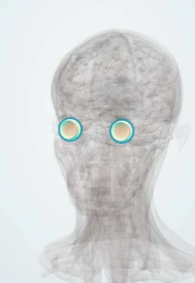
Who gets it
Rare; mostly boys in the first decade, usually one eye.
Causes
- Unknown; possible somatic mutations affecting retinal vessel development
Risk factors
- Male sex
- Young age
Pathophysiology
Abnormal, dilated retinal vessels with a broken blood-retinal barrier leak serum and lipid, which accumulate under and within the retina and lead to exudative detachment.
Classification
- By stage: telangiectasia only, exudation without or with macular involvement, partial or total exudative detachment, glaucoma, end-stage
Symptoms and signs
- Reduced vision in one eye
- Strabismus
- Yellow or white pupil reflex
Diagnosis
- Clinical
- Peripheral telangiectatic, light-bulb aneurysms with yellow subretinal exudate, usually in one eye.
- Tests
- Referral to paediatric ophthalmology; examination under anaesthesia in young children.
- Imaging
- Fluorescein angiography shows leaking telangiectasia; ultrasound shows exudative detachment without the calcification typical of retinoblastoma.
Differential diagnosis
Leukocoria in a child: Coats disease and its look-alikes
| Feature | Coats disease | Retinoblastoma | Familial exudative vitreoretinopathy |
|---|---|---|---|
| Typical patient | Boy in the first decade, one eye | Infant or toddler of either sex, one or both eyes | Either sex, both eyes, family history |
| Pupil reflex | Yellowish | White | Variable; white in severe cases |
| Fundus | Peripheral telangiectasia, light-bulb aneurysms, yellow exudate | White raised mass with feeding vessels, vitreous seeds | Avascular peripheral retina, dragged vessels, retinal folds |
| Ultrasound or CT | Exudative detachment without calcification | Mass with calcification | Traction and folds, no calcification |
| Inheritance | Not inherited | Germline RB1 mutation in heritable cases | Inherited, several genes |
Goal of treatment
Close the leaking vessels to dry the retina and preserve vision and the eye.
Treatment
- Procedure
- Laser photocoagulation or cryotherapy of the abnormal vessels.
- Medical
- Intravitreal anti-VEGF or corticosteroids as adjuncts in selected cases.
- Surgical
- Drainage of subretinal fluid and vitreoretinal surgery for advanced detachment; enucleation for a blind, painful eye.
Complications
- Exudative retinal detachment
- Neovascular glaucoma
- Amblyopia
- Loss of the eye
Common traps
- A boy with yellow exudate and leukocoria clearly has Coats disease: retinoblastoma must be excluded first, with an expert exam under anaesthesia and imaging, because missing it can be fatal.
- Calcification on ultrasound fits Coats disease: intraocular calcification in a child strongly suggests retinoblastoma; the exudate of Coats disease is usually not calcified.
- Coats disease is usually bilateral and inherited: it is typically one-eyed and sporadic; exudative vessel disease in both eyes with a family history suggests familial exudative vitreoretinopathy.
- A child with a new squint only needs patching: strabismus can be the first sign of a retinal disease such as Coats disease or retinoblastoma, so every child with new strabismus needs a dilated fundus exam.
- The yellow material is tumour tissue: it is lipid leaked from the abnormal vessels, and it is slowly reabsorbed once the vessels are closed.
Prognosis
Good when treated early; advanced disease often leaves poor vision.
Test yourself
Think of the answer, then tap the question to see it.
A 6-year-old boy has a yellowish pupil reflex and a squint in one eye. What must be excluded first, and how?
Retinoblastoma: urgent paediatric ophthalmology referral, examination under anaesthesia, and ultrasound or MRI looking for a mass and calcification.
Why does the retina detach in Coats disease?
Leaking abnormal vessels pour lipid-rich serum under the retina and lift it from below: an exudative detachment, not one caused by a retinal tear.
Why does laser or cryotherapy of the abnormal vessels help?
Closing the leaking vessels stops new exudate, so the existing fluid and lipid are slowly reabsorbed and the retina can flatten.
Why can advanced Coats disease cause a painful red eye?
A chronically detached, ischaemic retina drives new vessels on the iris and in the drainage angle, causing neovascular glaucoma.
In ophthalmology
Connections
Educational information, not medical advice. For any symptom, see a health professional.
Sources: Exudative Retinitis (Coats Disease) (StatPearls Publishing (NCBI Bookshelf), 2023).
Spotted a mistake?We are always improving for the community. Tell us what is wrong and our team will review it.Report a mistake
Representative model, for educational purposes only.