
Persistent fetal vasculature
A birth defect in which the eye's fetal blood vessels fail to regress, leaving a white pupil, a small eye and cataract. It is usually one-sided.
Quick facts
- Key signs
- White pupil · Small eye · Strabismus
- Diagnosis
- Axial length measurement
Red flags ↓
No doses: the clinician sets them by the current guideline.
What it is
Formerly called persistent hyperplastic primary vitreous: the hyaloid artery and the vessels around the fetal lens fail to regress before birth. It ranges from a harmless Mittendorf dot or Bergmeister papilla to a fibrovascular plaque behind the lens with stretched ciliary processes, cataract and a stalk to the disc.
Who gets it
Usually sporadic and unilateral (about 90% of cases) in otherwise healthy full-term infants. It is a leading non-cancerous cause of leukocoria; bilateral cases suggest a syndrome such as trisomy 13 or Norrie disease.
Causes
- Failure of the normal programmed regression of the hyaloid vascular system
- Mostly sporadic
- Bilateral cases: trisomy 13, Norrie disease, Walker-Warburg syndrome and other syndromes
Risk factors
- No consistent risk factors; bilateral disease calls for a genetic and systemic work-up
Pathophysiology
The hyaloid artery feeds the developing lens and vitreous and normally disappears by birth. When it persists, fibrovascular tissue behind the lens contracts, pulling the ciliary processes inward, clouding the lens and shallowing the anterior chamber; posterior forms tether the retina to a stalk, causing folds and detachment.
Classification
- Anterior: retrolental membrane, cataract, elongated ciliary processes, shallow anterior chamber
- Posterior: stalk from the disc, retinal fold, optic nerve hypoplasia or detachment
- Combined anterior and posterior (most common)
- Minor remnants: Mittendorf dot (back of the lens) and Bergmeister papilla (on the disc)
Symptoms and signs
- White pupil (leukocoria)
- Small eye (microphthalmia)
- Strabismus
- Poor vision in one eye
- Nystagmus in bilateral cases
Diagnosis
- Clinical
- A unilateral small eye with a retrolental plaque, elongated ciliary processes seen through the dilated pupil, cataract and a vascular stalk; check the red reflex in both eyes.
- Tests
- Examination under anaesthesia with intraocular pressure measurement; karyotype or genetic testing in bilateral cases.
- Imaging
- B-scan ultrasound shows a stalk from the disc to the back of the lens, without calcification; MRI helps exclude retinoblastoma when in doubt; axial length measurement confirms the small eye.
Differential diagnosis
- Retinoblastoma
- Congenital cataract
- Coats disease
- Retinopathy of prematurity
- Familial exudative vitreoretinopathy
- Ocular toxocariasis
- Norrie disease
White pupil in an infant: which one?
| Condition | Eye size | Key sign | Ultrasound | Laterality | Priority |
|---|---|---|---|---|---|
| Persistent fetal vasculature | Small | Plaque behind the lens, elongated ciliary processes, stalk to the disc | Stalk from disc to lens, no calcification | Usually one eye | Early surgery to save vision |
| Retinoblastoma | Normal | White or cream retinal mass, often with seeds | Solid mass with calcification | One or both eyes | Life-threatening; immediate referral |
| Congenital cataract | Usually normal | Lens opacity with a normal eye behind it | Normal posterior segment | One or both eyes | Early surgery to prevent amblyopia |
| Coats disease | Normal | Yellow exudate under dilated, leaking vessels | Exudative detachment, usually without calcification | Usually one eye, mostly boys | Laser or cryotherapy to stop exudation |
Goal of treatment
Exclude retinoblastoma, keep the eye and useful vision, and prevent glaucoma and amblyopia.
Treatment
- Surgical
- Early lensectomy and vitrectomy with removal of the retrolental membrane in infancy; posterior vitrectomy when there is traction.
- Rehabilitation
- Aphakic contact lens or glasses with patching of the better eye to treat amblyopia.
- Follow-up
- Observation of mild cases that spare the visual axis; lifelong pressure checks for glaucoma.
- Medical
- Pressure-lowering eye drops, and glaucoma surgery if needed, when pressure rises.
Complications
- Angle-closure or other secondary glaucoma
- Dense amblyopia
- Intraocular haemorrhage
- Retinal detachment
- Phthisis bulbi
Common traps
- Leukocoria in a small eye cannot be retinoblastoma, so imaging can wait: every white pupil needs prompt specialist examination and imaging, because only that excludes retinoblastoma
- PFV is usually bilateral and genetic: about 90% of cases are unilateral and sporadic in healthy term infants, and bilateral disease suggests a syndrome such as trisomy 13 or Norrie disease
- A Mittendorf dot or Bergmeister papilla needs treatment: they are harmless minor remnants of the same vascular system
- Once the cataract is removed the job is done: the eye still needs aphakic correction, patching against dense amblyopia and lifelong pressure checks for glaucoma
- PFV and retinoblastoma look the same on ultrasound: PFV shows a small eye with a stalk and no calcification, whereas retinoblastoma is a calcified mass in a normal-sized eye
Prognosis
Depends on severity: anterior forms treated early can reach useful vision, while posterior forms with retinal or optic nerve involvement have a poor visual outcome.
Prevention
- Red reflex test at birth and at every well-child visit
Test yourself
Think of the answer, then tap the question to see it.
A healthy term baby has a white pupil in a slightly small right eye. What must be done first?
Prompt specialist examination with ultrasound, and MRI if in doubt, to exclude retinoblastoma before accepting persistent fetal vasculature.
Why are the ciliary processes elongated in anterior PFV?
The plaque behind the lens is attached to them and contracts, pulling them inward so they become visible through the dilated pupil.
Why can angle-closure glaucoma develop in PFV?
The contracting membrane and a swollen or forward-moving lens shallow the anterior chamber and close the drainage angle.
Why do anterior forms have a better visual outcome than posterior forms?
Anterior disease is mainly lens and membrane, which surgery can clear; posterior disease brings retinal folds, detachment or optic nerve hypoplasia that cannot be restored.
A baby has PFV in both eyes. What extra work-up is needed?
Systemic and genetic assessment, including a karyotype, for syndromes such as trisomy 13, Norrie disease or Walker-Warburg syndrome.
Connections
Educational information, not medical advice. For any symptom, see a health professional.
Sources: Persistent Hyperplastic Primary Vitreous (EyeWiki (American Academy of Ophthalmology), 2025); Leukocoria (StatPearls Publishing (NCBI Bookshelf), 2023).
Spotted a mistake?We are always improving for the community. Tell us what is wrong and our team will review it.Report a mistake
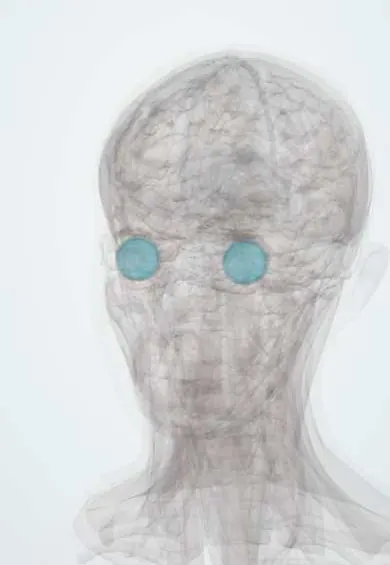
Representative model, for educational purposes only.