Conditions · Hematology and oncology
Disseminated intravascular coagulation !
DIC is a dangerous state in which clotting switches on throughout the body, using up platelets and clotting factors so that clots and bleeding occur together.
Updated 2026-11-01
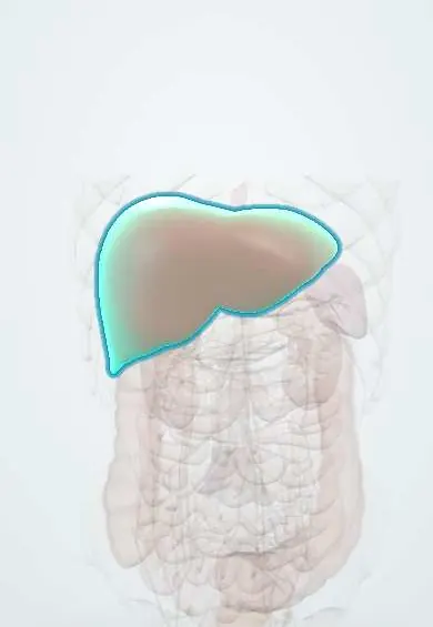
Structures involved
Tap one to see what it is and open it in 3D.
In the clinical exam
Why it happens
- Disseminated intravascular coagulation: how consumption causes both clots and bleeding
- How septic shock develops
- Thermal injury: burns, hyperthermia and hypothermia
- Fat, air and amniotic fluid embolism
- The coagulation cascade
- Fibrinolysis and natural anticoagulants
What it is
An acquired syndrome in which widespread activation of coagulation forms microthrombi in small vessels and consumes platelets and clotting factors. It is always secondary to another disease and can present as bleeding, organ damage from clots, or both.
Who gets it
Seen in a large share of patients with severe sepsis and septic shock, after major trauma and in obstetric emergencies. In Latin America, severe dengue and meningococcal disease are important infectious triggers, and Bothrops snakebite causes a consumption coagulopathy with a similar laboratory picture.
Causes
- Sepsis, the most common cause (gram-negative and gram-positive bacteria, meningococcaemia)
- Major trauma, burns and head injury
- Obstetric: placental abruption, amniotic fluid embolism, HELLP and preeclampsia, retained dead fetus, postpartum haemorrhage
- Cancer: acute promyelocytic leukaemia, mucin-producing adenocarcinomas (chronic DIC)
- Acute pancreatitis, severe liver failure, heat stroke
- Haemolytic transfusion reaction and some venoms
Risk factors
- Shock and prolonged hypotension
- Critical illness in an intensive care unit
- Late pregnancy and the peripartum period
- Active cancer, especially APL and metastatic adenocarcinoma
Pathophysiology
The trigger exposes tissue factor or releases procoagulants, so thrombin is generated throughout the circulation. Fibrin microthrombi block small vessels and injure kidney, lung, liver, brain and adrenals, while natural anticoagulants are used up. Platelets and factors are consumed faster than they are made, and fibrinolysis breaks fibrin into D-dimer, so the patient bleeds as well as clots. Red cells shear on fibrin strands, forming schistocytes.
Classification
- Acute (decompensated): bleeding predominates; sepsis, trauma, obstetric causes, APL
- Chronic (compensated): thrombosis predominates, labs may be near normal; solid cancers, retained dead fetus, large aneurysms
Symptoms and signs
- Oozing from venipuncture sites, catheters, wounds and gums
- Petechiae, purpura and ecchymoses
- Haematuria, gastrointestinal bleeding
- Signs of organ ischaemia: oliguria, confusion, breathlessness, cold or mottled fingers and toes
- Purpura fulminans (skin necrosis) in severe sepsis
Diagnosis
- Clinical
- Suspect it in a patient with a known trigger who bleeds from several sites or develops unexplained organ failure. Nurses monitor puncture sites, urine, stools, neurological status and vital signs closely.
- Tests
- Low or falling platelet count, prolonged prothrombin time and aPTT, low fibrinogen and markedly raised D-dimer or fibrin degradation products; schistocytes on the blood smear. Scoring systems such as the ISTH score combine these values. Serial testing matters more than a single result.
- Imaging
- Not diagnostic. Used to find the cause (e.g. obstetric ultrasound for abruption, CT for abdominal sepsis or pancreatitis) and to look for bleeding or thrombosis.
Differential diagnosis
- Thrombotic thrombocytopenic purpura and haemolytic uraemic syndrome (normal clotting times)
- HELLP syndrome
- Coagulopathy of liver disease (factor VIII usually normal or high)
- Vitamin K deficiency or anticoagulant excess
- Heparin-induced thrombocytopenia
- Dilutional coagulopathy after massive transfusion
Goal of treatment
Remove or treat the underlying cause as fast as possible, while replacing platelets and clotting factors only when the patient bleeds or needs a procedure, and preventing further organ damage.
Treatment
- Acute
- Treat the trigger: antibiotics and source control in sepsis, delivery of the fetus and placenta in obstetric causes, specific therapy for APL, antivenom in snakebite.
- Medical
- Blood components guided by bleeding and laboratory values: platelet transfusion, fresh frozen plasma, and cryoprecipitate or fibrinogen concentrate when fibrinogen is low.
- Medical
- Heparin in selected cases where thrombosis predominates (e.g. chronic DIC with cancer); antifibrinolytics are generally avoided.
- Follow-up
- Serial platelets, PT, aPTT, fibrinogen and D-dimer; organ support in intensive care; bleeding precautions (avoid intramuscular injections, apply pressure to puncture sites).
Complications
- Life-threatening haemorrhage, including intracranial
- Acute kidney injury
- Acute respiratory distress syndrome
- Hepatic dysfunction and adrenal infarction or haemorrhage
- Digital or limb gangrene
- Multi-organ failure and death
Prognosis
Prognosis depends mainly on the cause and its reversibility. Obstetric DIC often resolves quickly after delivery, while DIC in septic shock or major trauma carries high mortality.
Test yourself
Think of the answer, then tap the question to see it.
What is the most common cause of disseminated intravascular coagulation?
Sepsis.
Which laboratory pattern is typical of acute DIC?
Low platelets, prolonged PT and aPTT, low fibrinogen, high D-dimer and schistocytes on the smear.
How do TTP and DIC differ on coagulation tests?
Both lower platelets and cause schistocytes, but PT, aPTT and fibrinogen are usually normal in TTP and abnormal in DIC.
What is the cornerstone of DIC treatment?
Treating the underlying cause; blood components are given for bleeding or before procedures.
Which leukaemia classically presents with DIC?
Acute promyelocytic leukaemia (AML with t(15;17)).
Related conditions
- Sepsis and septic shock
- Acute leukaemia (AML and ALL)
- Placental abruption
- Preeclampsia
- Acute pancreatitis
- Acute kidney injury
- Acute respiratory distress syndrome (ARDS)
- Dengue
Educational information, not medical advice. For any symptom, see a health professional.
Sources: Disseminated intravascular coagulation (DIC) (MedlinePlus (U.S. National Library of Medicine), 2025).
Spotted a mistake?We are always improving for the community. Tell us what is wrong and our team will review it.Report a mistake
Representative model, for educational purposes only.