Conditions · Hematology and oncology
Acute leukaemia (AML and ALL) !
Acute leukaemia is a fast-growing blood cancer in which immature cells fill the bone marrow. ALL is the most common cancer of childhood.
Updated 2026-11-01
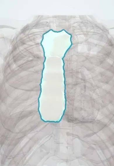
Structures involved
Tap one to see what it is and open it in 3D.
In embryology
In the clinical exam
Why it happens
- Translocations, amplifications and epigenetic changes in cancer
- Oncogenes: RAS, MYC, HER2, BCR-ABL and the table to know
- Ionizing radiation injury
- Trisomies 21, 18 and 13, and the 22q11.2 deletion
- Tumours of infancy and childhood
- Disseminated intravascular coagulation: how consumption causes both clots and bleeding
Show 8 more
- Chemical carcinogens: initiation, promotion and the table to know
- Radiation and cancer: ultraviolet and ionizing
- Cancer epidemiology: age, environment, predisposing conditions (and Colombia)
- Air pollution and occupational exposures
- Molecular pathology: PCR, FISH, sequencing and liquid biopsy
- Hematopoiesis
- Hematopoiesis and bone marrow
- Blood cells on a smear: erythrocytes, leukocytes, platelets
What it is
A group of blood cancers in which immature white cells (blasts) multiply in the bone marrow and crowd out normal blood production. Acute lymphoblastic leukaemia (ALL) arises from lymphoid precursors and acute myeloid leukaemia (AML) from myeloid precursors; both are fatal within weeks to months without treatment.
Who gets it
ALL is the leading cancer of childhood, with a peak between 2 and 5 years, and children of Hispanic background have some of the highest rates worldwide. AML is mainly a disease of older adults. In Colombia, leukaemias are the most frequent cancer in children and are under public-health surveillance; as in other middle-income countries, late diagnosis and treatment interruption lower survival compared with high-income settings.
Causes
- Acquired genetic changes in a blood-forming stem or precursor cell (translocations, mutations)
- Most cases have no identifiable cause
- Some arise after earlier chemotherapy or radiotherapy (therapy-related AML)
Risk factors
- Down syndrome and other inherited syndromes (Fanconi anaemia, Li-Fraumeni)
- Ionising radiation and previous chemotherapy (alkylating agents, topoisomerase II inhibitors)
- Benzene exposure and smoking (AML)
- Myelodysplastic syndrome or myeloproliferative neoplasm progressing to AML
- Age: ALL peaks in early childhood; AML rises with age
Pathophysiology
A clone of blasts that cannot mature accumulates in the marrow and spills into the blood. Normal production fails, causing anaemia, neutropenia and thrombocytopenia. Blasts infiltrate organs (liver, spleen, lymph nodes, gums, skin, central nervous system and testes in ALL). In acute promyelocytic leukaemia (APL), granules released by promyelocytes trigger disseminated intravascular coagulation, and very high blast counts can sludge small vessels (leukostasis).
Classification
- ALL: B-cell (most common, especially in children) or T-cell (often a mediastinal mass in adolescents)
- AML: classified by genetic abnormality (e.g. t(15;17) PML-RARA in APL, core-binding factor leukaemias) and by lineage
- Risk groups by age, white count, genetics and response to induction (e.g. Philadelphia chromosome t(9;22) is high risk in ALL)
Symptoms and signs
- Fatigue, pallor and breathlessness from anaemia
- Fever and recurrent infections from lack of functioning neutrophils
- Easy bruising, petechiae, gum bleeding and nosebleeds from low platelets
- Bone pain or limping in children; refusal to walk
- Enlarged liver, spleen or lymph nodes (more typical of ALL)
- Gum swelling or skin nodules (monocytic AML)
Diagnosis
- Clinical
- Consider it in any child or adult with unexplained cytopenias, fever, bleeding, bone pain or organ enlargement. Examine lymph nodes, liver, spleen, gums, skin and, in boys with ALL, the testes.
- Tests
- Complete blood count with a peripheral smear showing blasts; Auer rods point to AML. Bone marrow aspirate and biopsy with flow cytometry (immunophenotype), cytogenetics and molecular studies confirm the type; widely used criteria require about 20% or more blasts. Coagulation tests and fibrinogen (DIC), uric acid, potassium, phosphate, calcium, LDH and creatinine (tumour lysis), and cerebrospinal fluid examination in ALL.
- Imaging
- Chest X-ray or CT for a mediastinal mass, especially in T-cell ALL; echocardiography before anthracyclines; other imaging only for specific complications.
Differential diagnosis
- Aplastic anaemia
- Immune thrombocytopenia
- Infectious mononucleosis and other viral infections with atypical lymphocytes
- Myelodysplastic syndrome and chronic leukaemias
- Marrow infiltration by other cancers (neuroblastoma, lymphoma)
- Juvenile idiopathic arthritis in a child with bone or joint pain
Goal of treatment
Achieve complete remission with intensive chemotherapy, prevent relapse (including in the central nervous system), and support the patient through cytopenias, infection, bleeding and metabolic complications.
Treatment
- Medical
- Multi-agent chemotherapy in phases: induction to reach remission, then consolidation; ALL also receives central nervous system prophylaxis with intrathecal chemotherapy and a long maintenance phase.
- Medical
- Targeted treatment by subtype: all-trans retinoic acid with arsenic trioxide for APL, tyrosine kinase inhibitors for Philadelphia-positive ALL, and other targeted or immune therapies (monoclonal antibodies, CAR-T cells) in selected cases.
- Procedure
- Allogeneic haematopoietic stem cell transplantation for high-risk or relapsed disease.
- Acute
- Supportive care: irradiated blood products and platelet transfusions, hydration and uric acid-lowering drugs to prevent tumour lysis, urgent cytoreduction for leukostasis, and empirical antibiotics for febrile neutropenia.
- Follow-up
- Nursing priorities: neutropenic and bleeding precautions, oral care, daily weight and fluid balance, electrolytes and creatinine during induction, central line care, and minimal residual disease testing to guide therapy.
Complications
- Severe bacterial and fungal infections
- Haemorrhage, including intracranial bleeding
- Disseminated intravascular coagulation (classic in APL)
- Tumour lysis syndrome and acute kidney injury
- Leukostasis and superior vena cava syndrome
- Late effects of treatment: infertility, cardiotoxicity from anthracyclines, growth and learning problems, second cancers
Prognosis
Childhood ALL is cured in most children when full treatment is delivered without interruption. Adult ALL and AML have lower cure rates that fall with age and adverse genetics. APL, once treated promptly, has one of the best outcomes of any leukaemia, but early bleeding deaths remain a risk.
Test yourself
Think of the answer, then tap the question to see it.
What is the most common cancer in children, and at what age does it peak?
Acute lymphoblastic leukaemia, with a peak between about 2 and 5 years of age.
Which AML subtype is associated with t(15;17) and disseminated intravascular coagulation?
Acute promyelocytic leukaemia (APL), treated urgently with all-trans retinoic acid and arsenic trioxide.
Which finding on the peripheral smear points to AML rather than ALL?
Auer rods, needle-like inclusions in myeloid blasts.
Why does ALL treatment include intrathecal chemotherapy?
The central nervous system is a sanctuary site where blasts escape systemic chemotherapy, so it is treated directly to prevent relapse.
Which electrolyte pattern defines tumour lysis syndrome?
High potassium, phosphate and uric acid with low calcium, often with acute kidney injury.
Related conditions
- Febrile neutropenia
- Disseminated intravascular coagulation
- Tumour lysis syndrome
- Immune thrombocytopenia (ITP)
- Non-Hodgkin lymphoma
- Sepsis and septic shock
- Iron deficiency anaemia
Educational information, not medical advice. For any symptom, see a health professional.
Sources: Acute lymphoblastic leukemia (ALL) (MedlinePlus (U.S. National Library of Medicine), 2025); Acute myeloid leukemia - adult (MedlinePlus (U.S. National Library of Medicine), 2026); Childhood cancer (World Health Organization, 2026).
Spotted a mistake?We are always improving for the community. Tell us what is wrong and our team will review it.Report a mistake
Representative model, for educational purposes only.