Conditions · Metabolism (biochemistry)
Duchenne muscular dystrophy !
An inherited disease of boys in which dystrophin is missing: muscles are damaged and weaken from childhood.
Updated 2026-11-01
Structures involved
Tap one to see what it is and open it in 3D.
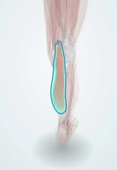
- Gastrocnemius (inner head)
- Gastrocnemius (outer head)
- Soleus
- Rectus femoris
- Vastus lateralis
- Gluteus maximus
- Diaphragm
- Left ventricle
In biochemistry
In the clinical exam
In physiology
- Sliding filaments and the cross-bridge cycle
- Motor units, recruitment and summation
- Muscle hypertrophy, atrophy and remodeling
Why it happens
- Mendelian inheritance: autosomal dominant, recessive and X-linked
- How a single gene causes disease: enzymes, receptors and structural proteins
What it is
An X-linked myopathy caused by DMD variants that abolish dystrophin, nearly always frameshifting deletions or nonsense mutations.
Who gets it
Duchenne and Becker together affect 1 in 3,500 to 5,000 newborn males worldwide; Duchenne is the most common childhood muscular dystrophy. About a third of cases are new mutations. No national registry has been published in Colombia.
Causes
- Variants in the DMD gene (Xp21), the largest human gene (79 exons)
- Deletions of one or more exons (≈ 65%), duplications (≈ 10%) and point mutations
- X-linked recessive inheritance; carrier mothers or a new mutation
Risk factors
- Male sex
- Carrier mother
- Affected male relatives on the mother's side
Pathophysiology
Dystrophin links the actin cytoskeleton to the membrane glycoprotein complex and the extracellular matrix; it acts as a shock absorber during contraction. If the deletion shifts the reading frame (frameshift), an early stop codon appears and no dystrophin is made (Duchenne). If the frame is kept, a shorter but working dystrophin is made (Becker, milder): this is the reading-frame rule. Without dystrophin the membrane tears with each contraction, calcium enters, proteases switch on and the fibre necroses; creatine kinase leaks into blood and muscle is replaced by fat and fibrous tissue (pseudohypertrophy).
Classification
- Duchenne: no dystrophin, onset at 2–5 years
- Becker: reduced or shorter dystrophin, later onset and slower course
- Female carriers: sometimes mild weakness or cardiomyopathy
Symptoms and signs
- Late walking, frequent falls, waddling gait and trouble running or climbing stairs
- Gowers sign: the child pushes on the legs with the hands to stand up from the floor
- Large, firm calves (pseudohypertrophy)
- Loss of walking around 10–13 years
- Scoliosis, contractures and, later, respiratory and heart failure
- Sometimes learning difficulties
Diagnosis
- Clinical
- A boy with proximal weakness, a Gowers sign and large calves.
- Tests
- Very high creatine kinase (tens of times normal); DMD genetic testing (MLPA for deletions and duplications, sequencing if negative); muscle biopsy showing absent dystrophin is kept for unclear cases.
- Imaging
- Muscle MRI shows the pattern of fatty infiltration; echocardiography and cardiac MRI follow the cardiomyopathy.
Treatment
- Medical
- Daily or intermittent corticosteroids (prednisone, deflazacort or vamorolone): slow the loss of strength.
- Medical
- Exon-skipping antisense oligonucleotides for eligible deletions (e.g. exon 51 or 53) and stop-codon readthrough drugs for nonsense mutations, where available.
- Medical
- Early cardioprotection (ACE inhibitors or ARBs, beta-blockers, mineralocorticoid antagonists).
- Procedure
- Night-time non-invasive ventilation and cough assistance when lung function falls.
- Surgical
- Spinal fusion for progressive scoliosis and release of selected contractures.
- Rehabilitation
- Physiotherapy, stretching, night splints and wheelchair fitting.
Complications
- Dilated cardiomyopathy and arrhythmias
- Restrictive respiratory failure
- Scoliosis
- Osteoporosis and fractures (worsened by corticosteroids)
- Rhabdomyolysis and hyperkalaemia with some anaesthetics
Prognosis
With corticosteroids, ventilation and cardiac care, survival has moved from adolescence to the third or fourth decade; cardiomyopathy is now the main cause of death.
Prevention
- Genetic counselling and carrier testing
- Prenatal or preimplantation diagnosis in affected families
Test yourself
Think of the answer, then tap the question to see it.
Why is Becker dystrophy milder than Duchenne?
In Becker the deletion keeps the reading frame and a short but working dystrophin is made; in Duchenne the frame shifts and there is no dystrophin.
What is the Gowers sign?
The child gets up from the floor by pushing the hands on the legs because of pelvic-girdle weakness.
Which first lab finding points to Duchenne?
A very high creatine kinase, tens of times the normal value.
How does exon skipping work?
An antisense oligonucleotide makes the spliceosome skip an exon next to the deletion and restores the reading frame.
Related conditions
Educational information, not medical advice. For any symptom, see a health professional.
Sources: Duchenne and Becker muscular dystrophy: MedlinePlus Genetics (MedlinePlus, US National Library of Medicine, 2026); DMD gene: MedlinePlus Genetics (MedlinePlus, US National Library of Medicine, 2026); Frameshift Mutation (National Human Genome Research Institute (NHGRI), 2026); Textbook of Biochemistry for Medical Students, 6th ed. (Jaypee Brothers Medical Publishers (Vasudevan DM, Sreekumari S, Vaidyanathan K), 2011).
Spotted a mistake?We are always improving for the community. Tell us what is wrong and our team will review it.Report a mistake
Representative model, for educational purposes only.