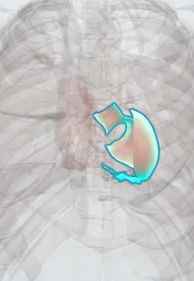
Left ventricle !
The left ventricle is the thick-walled lower left chamber of the heart; it pumps oxygen-rich blood through the aorta to the whole body.
Opens in the Durgical app, no sign-up needed.
What it is
The lower left chamber: the heart's main pump, with the thickest muscular wall (about 1 cm).
What it does
Pumps oxygen-rich blood through the aortic valve to the whole body, about 70 ml per beat at rest. Its pumping strength is measured as the ejection fraction (normally about 55-70%).
Key facts
- Where
- Forms apex, left border and most of diaphragmatic surface; wall about 3 times thicker than right ventricle
- Blood supply
- LAD (anterior wall, apex, anterior 2/3 of septum); circumflex (lateral wall); posterior descending (inferior wall)
- Nerve
- Cardiac plexus: sympathetic (T1–T4) speeds and strengthens; vagus nerves slow
- Function
- Pumps oxygenated blood through aortic valve into aorta at systemic pressure; apex beat at left 5th intercostal space
Relations
- In front
- Right ventricle and interventricular septum; along the left border a narrow strip of sternocostal surface, with the LAD (anterior descending artery) in the anterior interventricular groove
- Behind
- Left atrium above and behind, across the mitral valve; the circumflex artery and the coronary sinus in the atrioventricular groove
- Below
- Central tendon and left dome of the Diaphragm; the posterior descending artery (PDA) runs in the posterior interventricular groove
- Lateral
- Left lung and pleura across the pericardium, with the left phrenic nerve descending over it
- Above
- Aortic vestibule leading up to the aortic valve and the ascending aorta
- Contents
- Fine trabeculae carneae and the two Left ventricle papillary muscles, anchoring the mitral chordae tendineae
How to find it
The apex beat is felt with the fingertips in the left 5th intercostal space near the midclavicular line, more easily with the person turned onto the left side; in young children it lies a space higher. Mitral murmurs are heard there; its outflow is heard at the aortic area (right 2nd intercostal space).
On imaging
Frontal chest X-ray: the lower left heart border; enlargement raises the cardiothoracic ratio above about one half on a PA film and moves the apex down and out. Echo: parasternal and apical views measure wall thickness, cavity size, ejection fraction and regional wall motion, which maps to the coronary territories.
Common problems
- Heart attack: a blocked coronary artery kills part of its muscle.
- Heart failure: it pumps weakly (low ejection fraction) or becomes stiff and fills poorly.
- Thickens with long-standing high blood pressure or a narrowed aortic valve.
How it is diagnosed
ECG, blood tests (troponin) for a heart attack; echocardiogram for ejection fraction and wall movement; cardiac MRI.
Treatment
- Opening the blocked artery fast in a heart attack (angioplasty and stent).
- Heart-failure medicines, and devices such as a defibrillator or resynchronization pacemaker.
- Bypass surgery when several arteries are narrowed.
Clinical notes
- Mechanical complications of infarction: free wall rupture (tamponade), septal rupture (new harsh murmur) and papillary muscle rupture (acute mitral regurgitation)
- Left ventricular aneurysm after a large anterior infarct can hold thrombus and cause ventricular arrhythmias
- Long-standing hypertension or aortic stenosis thickens the wall (concentric hypertrophy); the ECG may show high voltage with a strain pattern
Anatomical variants
- False tendons (left ventricular bands): thin strands crossing the cavity, often from the septum to a papillary muscle; a common, usually harmless echo finding that can cause an innocent murmur
Development
It grows from the primitive ventricle of the heart tube, while the bulbus cordis builds most of the right ventricle. Its spongy embryonic wall normally compacts; if that fails, deep recesses between thick trabeculae remain (left ventricular non-compaction).
Related conditions
- Fluoroacetate poisoning
- Primary carnitine deficiency
- Beriberi (thiamine deficiency)
- Arterial hypertension
- Hypovolaemic shock
- Chagas disease
- Cardiac arrest (VF, pulseless VT, asystole, PEA)
- Hypertensive emergency and urgency
- Cardiogenic shock
- Ventricular tachycardia
- Hypertrophic cardiomyopathy
- Dilated cardiomyopathy
- Mechanical complications of myocardial infarction
- Myocarditis
- Wolff-Parkinson-White syndrome
- Long QT syndrome and torsades de pointes
- Syncope (vasovagal, orthostatic hypotension, cardiac)
- Transposition of the great arteries
- Takotsubo (stress) cardiomyopathy
- Peripartum cardiomyopathy
- Premature atrial and ventricular contractions
- Hypoplastic left heart syndrome
- Myocardial Infarction (Heart Attack)
- Aortic Valve Stenosis
- Heart failure
- Hypertensive heart disease
- Chagas cardiomyopathy
- Ventricular septal defect
Test yourself
Think of the answer, then tap the question to see it.
Which parts of the cardiac outline does the left ventricle form?
The apex, most of the left heart border on a frontal chest X-ray and most of the diaphragmatic surface.
Where is the apex beat felt, and what does a beat displaced down and out suggest?
In the left 5th intercostal space near the midclavicular line; displaced down and out, or broad, it suggests a dilated left ventricle.
What are the three mechanical complications of myocardial infarction involving the left ventricle?
Free wall rupture (tamponade), septal rupture (new harsh murmur) and papillary muscle rupture (acute mitral regurgitation).
Which left ventricular lesion is classic in Chagas cardiomyopathy?
A thin apical aneurysm, which can hold a thrombus.
Which coronary arteries supply the walls of the left ventricle?
LAD: anterior wall, apex and anterior 2/3 of the septum; circumflex: lateral wall; posterior descending artery: inferior wall.
Educational information, not medical advice. For any symptom, see a health professional.
Its names
- Official Latin (TA2)
- ventriculus sinister TA2 4062
- Official Spanish (DeCS)
- Ventrículo izquierdo DeCS D006352
- English (TA2)
- Left ventricle
Related
- Left ventricle papillary muscles
- Posterior vein of the left ventricle
- Left main coronary artery
- LAD (anterior descending artery)
- Circumflex artery
- Right coronary artery
- Posterior descending artery (PDA)
Sources: Terminologia Anatomica (TA2); DeCS; Anatomy, Thorax, Heart; Anatomy, Thorax, Heart and Pericardial Cavity; 19.5 Development of the Heart (OpenStax (Rice University), 2022) · adapted, CC BY 4.0; Tratado de anatomía humana (9.ª ed.), tomo II, Libro IV Angiología, sección 1, capítulo 1: Corazón, artículos I–VI.
Spotted a mistake?We are always improving for the community. Tell us what is wrong and our team will review it.Report a mistake
Representative model, for educational purposes only.