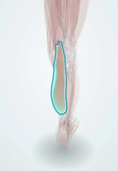
Gastrocnemius (inner head) !
The inner head of the gastrocnemius is the larger part of the calf muscle; it crosses the back of the knee and points the foot.
Official name: Medial head of gastrocnemius
Opens in the Durgical app, no sign-up needed.
What it is
The calf muscle (gastrocnemius). Its two heads start on the back of the femur and join the Achilles tendon.
What it does
Points the foot (pushing off when walking or jumping) and helps bend the knee.
Key facts
- Origin
- Posterior femur just above medial condyle
- Insertion
- Posterior calcaneus via calcaneal (Achilles) tendon
- Action
- Plantar flexes ankle; flexes knee
- Nerve
- Tibial nerve (S1–S2)
- Blood supply
- Medial sural artery (from popliteal artery)
Relations
- Above
- Its origin above the medial femoral condyle lies against the back of the knee capsule; between it and the semimembranosus tendon sits the gastrocnemius–semimembranosus bursa, which often opens into the knee joint
- Lateral
- The lateral head, separated by a midline groove that carries the small saphenous vein; deep in the gap between the heads, the popliteal vessels and tibial nerve pass down into the calf
- Deep
- Plantaris tendon and soleus; higher up, popliteus and the back of the knee capsule
- Medial
- Semimembranosus and the pes anserinus tendons at the knee; lower down, the great saphenous vein and saphenous nerve in the subcutaneous tissue of the medial leg
- Superficial
- Deep fascia and skin; the medial sural cutaneous nerve runs down over the heads and joins a branch of the common fibular nerve to form the sural nerve
How to find it
Standing on tiptoe, the medial head stands out as the larger, rounder bulge of the inner calf, its lower edge clearly lower than that of the lateral head. Lying prone with the knee bent against resistance, it is felt as the lower medial border of the popliteal fossa.
Examination
- Silfverskiöld test: ankle dorsiflexion is measured with the knee straight and then bent; if it is limited only with the knee straight the tightness is in the gastrocnemius, if limited in both positions the soleus or Achilles tendon is also tight
- Repeated single-leg heel raises: fewer or lower raises than on the other side point to calf weakness from a tear, a tibial nerve lesion or an S1 root problem
- Ankle reflex: a reduced or absent jerk suggests an S1 root or tibial nerve lesion (it is also often reduced in older people and in peripheral neuropathy)
On imaging
Ultrasound is the usual first test for tennis leg: a tear at the lower end of the medial head, where it meets the soleus, with fluid or haematoma between the two muscles. The same scan rules out a deep vein thrombosis, the main differential. A Baker's cyst bulges from the knee between this head and the semimembranosus tendon. MRI grades unclear tears.
Common problems
- Calf strain or tear, often in the inner head during a sudden push-off ('tennis leg').
- Cramps.
How it is diagnosed
Physical exam; ultrasound. Calf pain and swelling should also be checked for a blood clot.
Treatment
- Rest, ice, compression, then gradual stretching and strengthening.
Clinical notes
- Tennis leg typically follows a push-off with the knee straight and the ankle dorsiflexed; ultrasound shows the tear at the medial head with fluid between it and the soleus
- Popliteal artery entrapment: an abnormal course of the artery medial to or under the medial head, or an abnormal muscle slip, compresses it in young active people and causes exertional calf claudication
- Calf muscle pump: contraction squeezes the deep veins and the venous sinuses inside the calf muscles, driving blood upward through the valves; long immobility removes this pump and favours deep vein thrombosis
- A popliteal (Baker's) cyst bulges from the gastrocnemius–semimembranosus bursa; if it ruptures, fluid tracks down the calf and can mimic a deep vein thrombosis
- The medial head is the standard local muscle flap for exposed bone or hardware over the knee and upper third of the tibia; gastrocnemius recession lengthens an isolated gastrocnemius contracture
Anatomical variants
- A third head (gastrocnemius tertius) or an extra slip from the femur may be present and is one cause of popliteal artery entrapment
Development
The calf muscles come from the ventral (flexor) muscle mass of the leg, so the whole superficial posterior compartment is supplied by the tibial nerve.
Related conditions
- McArdle disease (glycogen storage disease type V)
- Carnitine palmitoyltransferase II (CPT-II) deficiency
- Duchenne muscular dystrophy
- Achilles tendon rupture
- Falls in older adults
- Frailty syndrome
- Sarcopenia
- Necrotizing soft tissue infection (necrotizing fasciitis, gas gangrene)
- Trichinellosis
- Rhabdomyolysis
Test yourself
Think of the answer, then tap the question to see it.
What is «tennis leg» and which muscle is usually torn?
A calf tear on push-off with the knee straight and ankle dorsiflexed; it is usually the medial head of the gastrocnemius, where it joins the soleus aponeurosis.
What does the Silfverskiöld test show?
If dorsiflexion is limited only with the knee straight the tightness is in the gastrocnemius; if limited with the knee straight and bent, the soleus or Achilles tendon is also tight.
Which nerve supplies the gastrocnemius, and which reflex tests it with the soleus?
The tibial nerve (S1–S2); the ankle jerk (S1) is the stretch reflex of the triceps surae (gastrocnemius and soleus).
Which border of the popliteal fossa does the medial head of the gastrocnemius form?
The inferomedial border; the lateral head and plantaris form the inferolateral border.
How does the medial head of the gastrocnemius cause popliteal artery entrapment?
An abnormal course of the artery medial to or under the medial head compresses it in young active people, causing exertional calf claudication.
Educational information, not medical advice. For any symptom, see a health professional.
Its names
- Official Latin (TA2)
- caput mediale musculi gastrocnemii TA2 2659
- Official Spanish (DeCS)
- Cabeza medial del músculo gastrocnemio
- English (TA2)
- Medial head of gastrocnemius
- Also called
- caput tibiale musculi gastrocnemii
Related
- Gastrocnemius (outer head)
- Soleus
- Tibial nerve
- Popliteal artery
- Gastrocnemius tendon (inner head)
- Femur
- Tibia
- Achilles tendon
Sources: Terminologia Anatomica (TA2); Anatomy, Bony Pelvis and Lower Limb: Gastrocnemius Muscle.
Spotted a mistake?We are always improving for the community. Tell us what is wrong and our team will review it.Report a mistake
Representative model, for educational purposes only.