
Ectopia lentis and lens shape anomalies
The lens slips out of place because its support fibres are weak, often in Marfan syndrome or homocystinuria. Vision blurs and shifts.
What it is
Displacement of the lens because its zonular fibres are weak or broken, either partial (subluxation) or complete (luxation). Causes are trauma or inherited conditions such as Marfan syndrome and homocystinuria. Related lens shape anomalies include microspherophakia and lenticonus.
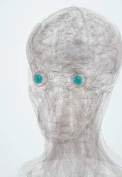
See it in 3D
Who gets it
Trauma is the commonest cause overall. Among inherited causes, Marfan syndrome is the most frequent; ectopia lentis occurs in most people with Marfan syndrome and most untreated people with classic homocystinuria.
Causes
- Trauma (blunt injury)
- Marfan syndrome (FBN1, fibrillin-1): usually upward and outward displacement
- Homocystinuria (CBS deficiency): usually downward and inward
- Weill-Marchesani syndrome (microspherophakia, short stature), sulfite oxidase deficiency, isolated familial ectopia lentis
- Pseudoexfoliation, high myopia, buphthalmos, hypermature cataract
- Lenticonus: anterior in Alport syndrome (COL4A5), posterior usually isolated
Risk factors
- Connective tissue and metabolic disorders
- Blunt eye trauma
- Family history
Pathophysiology
Zonular fibres are built on fibrillin microfibrils. Faulty fibrillin (Marfan) or disrupted disulfide bonds from excess homocysteine stretch or break them, so the lens shifts away from the weakest zonules. A displaced, rounder lens causes large refractive changes and, if it moves forward or into the pupil, blocks aqueous flow and causes angle-closure glaucoma.
Classification
- Subluxation (partly displaced, still behind the iris) or luxation (into the vitreous or anterior chamber)
- Traumatic or hereditary (with or without systemic disease)
- Microspherophakia: small, spherical lens with high myopia and pupil block risk
- Lenticonus: conical bulge of the anterior or posterior lens surface
Symptoms and signs
- Blurred or fluctuating vision, often high myopia or astigmatism
- Monocular double vision when the lens edge crosses the pupil
- Trembling of the iris (iridodonesis) or lens (phacodonesis)
- Painful red eye and halos if pupil block glaucoma develops
- Signs of the systemic condition (tall stature, long limbs, intellectual disability, thromboses)
Diagnosis
- Clinical
- Slit-lamp examination with the pupil dilated to see the lens edge and direction of displacement; refraction; eye pressure and gonioscopy; systemic examination for Marfan features.
- Tests
- Plasma total homocysteine and amino acids for homocystinuria; FBN1 and other genetic testing; urinalysis and hearing tests for Alport syndrome.
- Imaging
- Ultrasound biomicroscopy for zonular and lens assessment; B-scan ultrasound for a lens in the vitreous; echocardiography for the aortic root in Marfan syndrome.
Differential diagnosis
- Pseudoexfoliation with zonular weakness
- Traumatic cataract
- Iris coloboma (mimicking a lens notch)
- Keratoconus as a cause of high astigmatism
Ectopia lentis: the main causes compared
| Feature | Marfan syndrome | Homocystinuria | Weill-Marchesani syndrome | Trauma |
|---|---|---|---|---|
| Defect | Fibrillin-1 (FBN1) | Cystathionine beta-synthase deficiency; homocysteine damages the zonules | Small, spherical lens (microspherophakia) | Zonules torn by blunt injury |
| Usual direction | Up and out | Down and in | Forward, into the pupil | Any direction, usually one eye |
| Build | Tall, long limbs, lax joints | Tall, often intellectual disability | Short stature, short fingers, stiff joints | Unremarkable |
| Main danger beyond blur | Aortic root dilatation and dissection | Thromboembolism, especially with anaesthesia | Pupil block glaucoma | Associated injuries (hyphema, retinal detachment) |
| Key test | Echocardiography, FBN1 testing | Plasma total homocysteine | Clinical features, genetic testing | History and full eye examination |
Goal of treatment
Give the clearest possible vision, prevent amblyopia in children and avoid glaucoma and retinal complications, while managing the systemic disease.
Treatment
- Conservative
- Glasses or contact lenses correcting through the phakic or aphakic part of the pupil, with amblyopia treatment in children.
- Medical
- For homocystinuria: pyridoxine trial, low-methionine diet, betaine and folate with vitamin B12 under metabolic specialists; for pupil block: laser iridotomy and pressure-lowering treatment.
- Surgical
- Lens removal (lensectomy or extraction with capsular tension devices) when vision cannot be corrected, with an iris- or scleral-fixated intraocular lens or aphakic correction; urgent removal of a lens in the anterior chamber.
- Follow-up
- Lifelong eye follow-up for glaucoma and retinal detachment; cardiology follow-up in Marfan syndrome.
Complications
- Amblyopia in children
- Pupil block and angle-closure glaucoma
- Retinal detachment
- Uveitis and corneal decompensation from a lens in the anterior chamber
- Thromboembolism during anaesthesia in homocystinuria
Common traps
- Lens displacement in a tall teenager is always Marfan syndrome: homocystinuria also gives tall stature and ectopia lentis, but with intellectual disability, thromboses and usually downward displacement, so plasma homocysteine is checked
- Homocystinuria and Marfan syndrome displace the lens the same way: in Marfan syndrome the lens usually shifts up and out, in homocystinuria down and in
- A child with ectopia lentis can go to theatre like any other child: undiagnosed homocystinuria carries a high risk of thrombosis under general anaesthesia, so homocysteine is measured first
- A displaced lens only affects sharpness of vision: a lens moving forward or into the pupil causes pupil block angle-closure glaucoma, an emergency
- Ectopia lentis is just an eye finding in Marfan syndrome: the same fibrillin defect weakens the aortic root, so echocardiography and cardiology follow-up are part of care
Prognosis
With early optical correction or surgery most children reach useful vision. Long-term outlook depends on retinal health and on the systemic disease, especially aortic disease in Marfan syndrome.
Prevention
- Newborn screening for homocystinuria where available
- Eye protection after blunt trauma and in connective tissue disorders
Test yourself
Think of the answer, then tap the question to see it.
A tall 12-year-old with learning difficulty has a lens displaced down and in, and her uncle had a stroke at 30. Which test comes first?
Plasma total homocysteine: the picture suggests homocystinuria, which also carries a high thrombosis risk during anaesthesia.
Why does the iris tremble with eye movements in ectopia lentis?
The iris normally rests on the lens; when the lens shifts, part of the iris loses its support and quivers (iridodonesis).
A patient with a subluxated lens suddenly develops a painful red eye with halos. What has happened?
The lens has moved forward and blocked the pupil, causing acute angle-closure glaucoma: an emergency needing pressure lowering, laser iridotomy or lens removal.
Why does a patient with Marfan syndrome and ectopia lentis need an echocardiogram?
The same fibrillin-1 defect weakens the aortic root, which can dilate and dissect.
A young adult has a small, spherical lens, high myopia, short stature and short fingers. Which syndrome is this?
Weill-Marchesani syndrome (microspherophakia).
Connections
In biochemistry1
Educational information, not medical advice. For any symptom, see a health professional.
Sources: Ectopia Lentis (StatPearls Publishing (NCBI Bookshelf), 2023); Microspherophakia (StatPearls Publishing (NCBI Bookshelf), 2026); Ectopia Lentis (EyeWiki, American Academy of Ophthalmology, 2026); Homocystinuria: MedlinePlus Genetics (MedlinePlus, US National Library of Medicine, 2023).
Spotted a mistake?We are always improving for the community. Tell us what is wrong and our team will review it.Report a mistake
Representative model, for educational purposes only.