Narcolepsy !
A lifelong sleep disorder causing overwhelming daytime sleepiness and, in type 1, sudden muscle weakness triggered by emotions (cataplexy).
Updated 2026-11-01
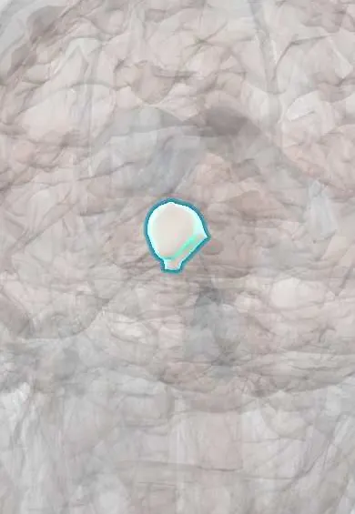
Structures involved
Tap one to see what it is and open it in 3D.
Why it happens
What it is
A chronic neurological disorder of the brain systems that control sleep and wakefulness. Its hallmark is excessive daytime sleepiness with irresistible sleep attacks. In type 1 there is cataplexy and loss of hypocretin (orexin) neurons in the hypothalamus; REM sleep intrudes into wakefulness, causing sleep paralysis and vivid hallucinations at sleep onset or awakening.
Who gets it
Affects about 1 in 2,000 people; onset is usually in adolescence or young adulthood, with a second peak in the 30s. Diagnosis is often delayed by 8-10 years because sleepiness is attributed to laziness, depression or poor sleep habits; in Latin America, limited access to sleep laboratories adds to the delay.
Causes
- Type 1: loss of hypocretin (orexin)-producing neurons in the lateral hypothalamus, thought to be autoimmune
- Strong genetic association with HLA-DQB1*06:02
- Possible triggers: streptococcal infection and influenza A (H1N1) infection or vaccination
- Secondary narcolepsy from hypothalamic lesions: tumours, trauma, stroke, inflammation
- Type 2: cause unknown, hypocretin usually normal
Risk factors
- HLA-DQB1*06:02 carriage
- First-degree relative with narcolepsy
- Recent streptococcal or influenza infection
- Hypothalamic injury
Pathophysiology
Hypocretin neurons in the lateral hypothalamus stabilise wakefulness and keep REM sleep switched off during the day by activating arousal centres in the brainstem, hypothalamus and basal forebrain. Their loss makes the sleep-wake switch unstable: patients fall asleep many times a day and enter REM sleep abnormally fast. Fragments of REM sleep appear during wakefulness: muscle atonia (from the pons and medulla) triggered by emotion is cataplexy; atonia at sleep transitions is sleep paralysis; dream imagery causes hypnagogic and hypnopompic hallucinations.
Classification
- Narcolepsy type 1: cataplexy and/or low CSF hypocretin-1
- Narcolepsy type 2: no cataplexy, normal hypocretin, positive sleep tests
- Secondary narcolepsy due to a hypothalamic lesion
Symptoms and signs
- Excessive daytime sleepiness with sudden sleep attacks, even while talking or eating; short naps are refreshing
- Cataplexy (type 1): brief sudden loss of muscle tone with preserved consciousness, triggered by laughter, surprise or anger (jaw drop, head drop, knee buckling)
- Sleep paralysis on falling asleep or waking
- Hypnagogic and hypnopompic hallucinations
- Fragmented night-time sleep
- Automatic behaviours, memory and attention problems; REM sleep behaviour disorder and weight gain in some
Diagnosis
- Clinical
- Suspected with daytime sleepiness for at least 3 months, especially with cataplexy. Sleepiness scales (Epworth) and a sleep diary help, and other causes such as insufficient sleep, sleep apnoea, depression and sedating drugs must be excluded.
- Tests
- Overnight polysomnography followed by a multiple sleep latency test (MSLT): mean sleep latency of 8 minutes or less with two or more sleep-onset REM periods. Low CSF hypocretin-1 confirms type 1. HLA typing supports but does not make the diagnosis.
- Imaging
- Brain MRI when secondary narcolepsy is suspected (hypothalamic lesion) or neurological signs are present.
Differential diagnosis
- Insufficient sleep and circadian rhythm disorders
- Obstructive sleep apnoea
- Idiopathic hypersomnia
- Depression and sedating medicines or substance use
- Epilepsy (atonic seizures) or syncope, in the case of cataplexy
- Psychosis, in the case of hallucinations
Goal of treatment
There is no cure; the goals are to keep the patient alert and safe during the day, control cataplexy and other REM symptoms and support school, work and driving safety.
Treatment
- Lifestyle
- Regular sleep schedule with planned short daytime naps, avoidance of alcohol and sedatives, and education for the patient, family, school or employer.
- Medical
- Wake-promoting agents (modafinil, armodafinil, pitolisant, solriamfetol) or stimulants (methylphenidate, amphetamines) for daytime sleepiness.
- Medical
- Sodium oxybate (and related oxybate salts) improves both sleepiness and cataplexy; antidepressants that suppress REM sleep (venlafaxine, SSRIs, tricyclics) treat cataplexy. Hypocretin (orexin) receptor agonists are an emerging class for type 1, still being evaluated.
- Follow-up
- Follow-up of sleepiness scores, cataplexy frequency, mood, blood pressure and heart rate on stimulants, and fitness to drive.
Complications
- Road traffic and work accidents
- Poor school or work performance
- Depression and anxiety
- Obesity and metabolic problems
- Social isolation and stigma
Prognosis
A lifelong condition that does not shorten life on its own. Symptoms usually stabilise after the first years, and with treatment most people can study, work and drive safely, although sleepiness rarely disappears completely.
Test yourself
Think of the answer, then tap the question to see it.
What neurotransmitter is lost in narcolepsy type 1, and where are the neurons?
Hypocretin (orexin), produced by neurons in the lateral hypothalamus.
What is cataplexy?
Sudden brief loss of muscle tone with preserved consciousness, triggered by strong emotion such as laughter.
Which sleep study findings confirm narcolepsy?
On the multiple sleep latency test, mean sleep latency of 8 minutes or less and at least two sleep-onset REM periods (after a normal overnight polysomnography).
What is the classic symptom tetrad?
Excessive daytime sleepiness, cataplexy, sleep paralysis and hypnagogic hallucinations.
Which drug treats both sleepiness and cataplexy?
Sodium oxybate.
Related conditions
- Obstructive sleep apnoea
- Insomnia disorder
- Epilepsy and seizures
- Syncope (vasovagal, orthostatic hypotension, cardiac)
- Major depressive disorder
- Attention-deficit/hyperactivity disorder (ADHD)
- Schizophrenia
- Dementia with Lewy bodies
Educational information, not medical advice. For any symptom, see a health professional.
Sources: Narcolepsy | National Institute of Neurological Disorders and Stroke (National Institute of Neurological Disorders and Stroke (NINDS), 2026).
Spotted a mistake?We are always improving for the community. Tell us what is wrong and our team will review it.Report a mistake
Representative model, for educational purposes only.