Conditions · Metabolism: carbohydrates and glycogen
Pompe disease (glycogen storage disease type II) !
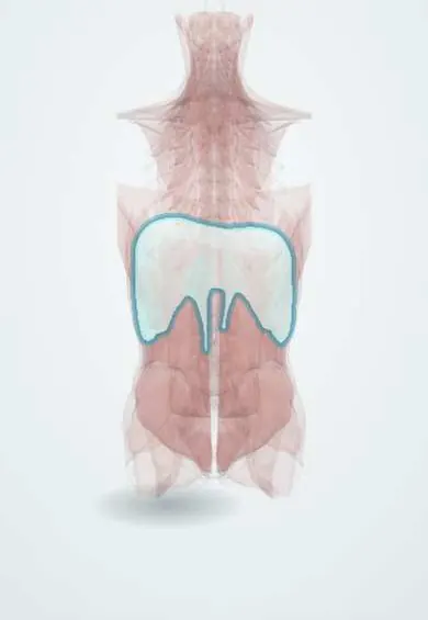
In Pompe disease glycogen builds up inside lysosomes and weakens the heart and muscles, including the diaphragm.
Updated 2026-11-01
Structures involved
Tap one to see what it is and open it in 3D.
In biochemistry
Why it happens
- Intracellular accumulations: cholesterol, proteins, hyaline, glycogen
- Autophagy
- How a single gene causes disease: enzymes, receptors and structural proteins
What it is
Lysosomal glycogen storage disease caused by lack of acid α-glucosidase (acid maltase, GAA gene). It is both a glycogen storage disease and a lysosomal storage disease.
Who gets it
About 1 in 40,000 people; frequency varies between populations. In Colombia and Latin America it is increasingly diagnosed thanks to dried blood spot testing.
Causes
- Autosomal recessive inheritance: GAA gene variants
Risk factors
- Carrier parents or affected siblings
Pathophysiology
A small fraction of glycogen enters lysosomes, where acid α-glucosidase breaks it down to glucose. Without it, glycogen fills the lysosomes, which burst and damage the fibres of heart, skeletal muscle and diaphragm. Blood glucose is normal because the cytosolic glycogen pathway works. The less residual activity, the earlier and more severe the form.
Classification
- Classic infantile: hypertrophic cardiomyopathy and hypotonia in the first months
- Non-classic infantile: weakness in the first year, with less heart involvement
- Late-onset (child or adult): proximal myopathy and respiratory failure, without cardiomyopathy
Symptoms and signs
- Hypotonic («floppy») baby who cannot hold up the head
- Large tongue and feeding difficulty
- Enlarged heart with heart failure
- In the late form: hip and thigh weakness, difficulty climbing stairs
- Breathlessness lying down and daytime sleepiness from diaphragm weakness
Diagnosis
- Clinical
- Hypotonic infant with macroglossia and cardiomegaly, or adult with limb-girdle myopathy and diaphragm weakness.
- Tests
- Raised CK; low acid α-glucosidase activity in dried blood spot or leukocytes; GAA gene testing; urinary glucose tetrasaccharide. ECG with short PR and very tall QRS in the infantile form. Spirometry sitting and lying in the late form.
- Imaging
- Chest X-ray with cardiomegaly; echocardiogram with hypertrophic cardiomyopathy; muscle MRI to see the affected muscles.
Treatment
- Medical
- Enzyme replacement therapy with recombinant acid α-glucosidase, started as early as possible
- Procedure
- Night-time non-invasive ventilation when the diaphragm weakens
- Rehabilitation
- Physiotherapy, respiratory therapy and speech therapy for swallowing
- Follow-up
- Cardiac, respiratory and muscle strength monitoring
Complications
- Heart failure and arrhythmias in the infantile form
- Respiratory failure
- Aspiration pneumonia
- Scoliosis
Prognosis
Untreated, the classic infantile form is usually fatal within the first year; early enzyme replacement greatly improves survival. The late form progresses slowly.
Test yourself
Think of the answer, then tap the question to see it.
Why does Pompe disease not cause hypoglycaemia?
The missing enzyme is lysosomal; cytosolic glycogen breakdown, which maintains blood glucose, works.
Which organ defines the severity of the classic infantile form?
The heart: hypertrophic cardiomyopathy with heart failure.
Which enzyme is missing in Pompe disease?
Acid α-glucosidase (acid maltase), encoded by GAA.
Which muscle explains respiratory failure in the late form?
The diaphragm; breathlessness worsens lying down.
Related conditions
- Heart failure
- McArdle disease (glycogen storage disease type V)
- Cori disease (glycogen storage disease type III)
Educational information, not medical advice. For any symptom, see a health professional.
Sources: Pompe disease: MedlinePlus Genetics (MedlinePlus, US National Library of Medicine, 2026); GAA gene: MedlinePlus Genetics (MedlinePlus, US National Library of Medicine, 2026); Textbook of Biochemistry for Medical Students, 6th ed. (Jaypee Brothers Medical Publishers (Vasudevan DM, Sreekumari S, Vaidyanathan K), 2011).
Spotted a mistake?We are always improving for the community. Tell us what is wrong and our team will review it.Report a mistake
Representative model, for educational purposes only.