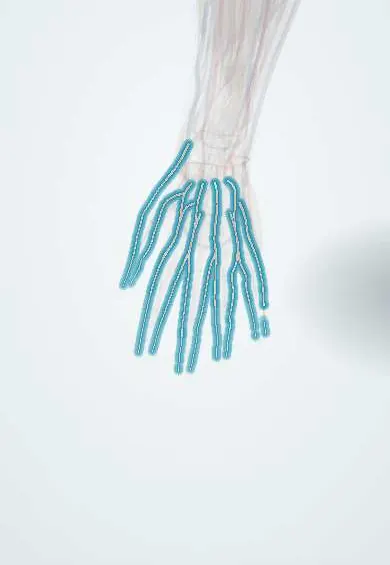
Raynaud phenomenon !
Raynaud phenomenon is attacks of narrowing of the small finger arteries with cold or stress, so the fingers turn white, then blue, then red.
Updated 2026-10-29
Structures involved
Tap one to see what it is and open it in 3D.
- Digital arteries
- Superficial palmar arch
- Deep palmar arch
- Radial artery
- Ulnar artery
- Subclavian artery
- Dorsal hand arteries
In the clinical exam
In physiology
Why it happens
- Adrenergic and cholinergic receptors
- Local and humoral control of blood flow
- Blood vessels: artery, vein and capillary layers
What it is
Episodes of exaggerated narrowing of the small arteries of the fingers (and sometimes toes, ears or nose) triggered by cold or emotional stress, which make the digits turn white, then blue and then red. It is primary when no cause is found and secondary when it accompanies another disease, most often systemic sclerosis.
Who gets it
Primary Raynaud is common, mainly in young women, often starting in the teens or twenties, and is more frequent in cold climates such as the Andean highlands. Secondary Raynaud appears later and occurs in nearly all patients with systemic sclerosis and in many with lupus.
Causes
- Primary (Raynaud disease): no underlying disease
- Connective tissue diseases: systemic sclerosis, systemic lupus erythematosus, Sjögren syndrome, dermatomyositis, mixed connective tissue disease
- Hand-arm vibration from tools, and repetitive trauma to the palm
- Drugs: beta-blockers, ergot derivatives, some chemotherapy agents and stimulants
- Arterial disease, thoracic outlet syndrome, cryoglobulinaemia and hyperviscosity states
Risk factors
- Female sex and young age (primary)
- Family history
- Cold exposure
- Smoking
- Connective tissue disease
- Work with vibrating tools
Pathophysiology
Cold and sympathetic stimulation cause an exaggerated alpha-adrenergic constriction of the digital arteries and arterioles. In primary Raynaud the vessels are structurally normal and the response is reversible. In secondary Raynaud endothelial damage, intimal thickening and microvascular loss make attacks more severe and can cause tissue ischaemia, ulcers and gangrene.
Classification
- Primary (Raynaud disease)
- Secondary (Raynaud phenomenon associated with another disease, drug or exposure)
- Colour phases: white (ischaemia), blue (deoxygenation), red (reactive hyperaemia on rewarming)
Symptoms and signs
- Well-demarcated whitening of one or more fingers on cold exposure or stress
- Blue discolouration followed by redness, throbbing and tingling on rewarming
- Numbness, clumsiness and pain during attacks
- Usually symmetric in primary Raynaud
- In secondary forms: fingertip ulcers, pitting scars, thickened skin or swollen fingers
Diagnosis
- Clinical
- History of episodic, cold-triggered colour changes with at least two phases, examination of the skin, nails and pulses, and a search for features of connective tissue disease. Criteria for primary Raynaud vary but include symmetric attacks, no ulcers, normal capillaries and negative antinuclear antibodies.
- Tests
- Antinuclear antibodies and, if positive, specific antibodies (anticentromere, anti-topoisomerase I); blood count, ESR and CRP; nailfold capillaroscopy.
- Imaging
- Not routine; Doppler ultrasound or angiography when large-artery disease or thoracic outlet compression is suspected.
Differential diagnosis
- Acrocyanosis
- Peripheral artery disease or embolism to the hand
- Thoracic outlet syndrome
- Chilblains (pernio)
- Hand-arm vibration syndrome
Goal of treatment
Reduce the frequency and severity of attacks, protect the fingers from ischaemic injury, and identify and treat any underlying disease.
Treatment
- Lifestyle
- Keeping the whole body and hands warm (gloves, layers), avoiding sudden cold and stress triggers, stopping smoking and avoiding vasoconstrictor drugs when possible.
- Medical
- Dihydropyridine calcium channel blockers are the usual first drug choice for frequent or troublesome attacks.
- Medical
- Further options include phosphodiesterase-5 inhibitors, topical nitrates and, for severe disease or digital ulcers, intravenous prostacyclin analogues; endothelin receptor antagonists are used to prevent new ulcers in systemic sclerosis.
- Procedure
- Botulinum toxin injection around the digital arteries in refractory cases.
- Surgical
- Digital or palmar sympathectomy for severe refractory ischaemia; debridement or amputation for gangrene.
Complications
- Digital ulcers and pitting scars
- Infection of ulcers and osteomyelitis
- Gangrene and loss of fingertips
- Reduced hand function and quality of life
Prognosis
Primary Raynaud is usually benign and may improve with age. A minority later develop a connective tissue disease, more often if capillaroscopy or autoantibodies are abnormal. Secondary Raynaud tends to be more severe and follows the course of the underlying disease.
Prevention
- Protection from cold
- Not smoking
- Limiting exposure to vibrating tools
Test yourself
Think of the answer, then tap the question to see it.
What are the three colour phases of a Raynaud attack?
White (ischaemia), blue (deoxygenation) and red (reactive hyperaemia on rewarming).
Which disease is most strongly associated with secondary Raynaud phenomenon?
Systemic sclerosis (scleroderma), in which Raynaud is present in nearly all patients.
Which features suggest secondary rather than primary Raynaud?
Onset later in life, asymmetric attacks, digital ulcers, abnormal nailfold capillaries, positive autoantibodies or signs of connective tissue disease.
What drug class is first-line for troublesome Raynaud phenomenon?
Dihydropyridine calcium channel blockers.
Which simple bedside test helps separate primary from secondary Raynaud?
Nailfold capillaroscopy: dilated, giant or lost capillaries suggest a connective tissue disease.
Related conditions
Educational information, not medical advice. For any symptom, see a health professional.
Sources: Raynaud Disease (StatPearls (NCBI Bookshelf), 2026).
Spotted a mistake?We are always improving for the community. Tell us what is wrong and our team will review it.Report a mistake
Representative model, for educational purposes only.