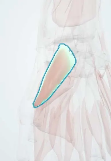
APB !
The abductor pollicis brevis is a thumb-pad muscle at the base of the thumb; it lifts the thumb away from the palm and wastes in severe carpal tunnel syndrome.
Official name: Abductor pollicis brevis
Opens in the Durgical app, no sign-up needed.
What it is
The surface muscle of the thumb pad (thenar eminence).
What it does
Lifts the thumb away from the palm; it wastes in severe carpal tunnel syndrome.
Key facts
- Origin
- Flexor retinaculum, tubercles of the scaphoid and trapezium
- Insertion
- Lateral base of the proximal phalanx of the thumb
- Action
- Abducts the thumb (palmar abduction)
- Nerve
- Recurrent (thenar) branch of the median nerve (C8–T1)
- Blood supply
- Superficial palmar branch of the radial artery
Relations
- Superficial
- Thin thenar fascia and skin, supplied by the palmar cutaneous branch of the median nerve
- Deep
- Opponens pollicis
- Medial
- Superficial head of flexor pollicis brevis (FPB)
- Above
- Its origin from the flexor retinaculum and the tubercles of the scaphoid and trapezium; the recurrent motor branch arrives here
How to find it
With the back of the hand on a table, raising the thumb straight up toward the ceiling against resistance makes it the firm outer bulge of the thenar eminence.
Examination
- Palmar abduction (pen) test: the thumb is raised to touch a pen held above the palm; weakness or a flat outer thenar eminence suggests a median nerve lesion at the wrist or above
On imaging
Not imaged on its own for diagnosis: it is the thenar muscle checked for wasting in carpal tunnel syndrome. Ultrasound assesses the median nerve, swollen at the carpal tunnel inlet, and can show thin thenar muscles; MRI shows denervation as oedema, later fatty atrophy. Nerve conduction studies remain the reference test.
Clinical notes
- Thenar wasting in carpal tunnel syndrome is seen first and most clearly in this muscle, a sign of advanced compression
- Opponensplasty transfers (ring finger FDS, extensor indicis, palmaris longus, abductor digiti minimi) are anchored at its insertion
- The recurrent motor branch can leave the median nerve above, below or through the flexor retinaculum, a known risk in carpal tunnel release
Development
A ventral mass muscle; the outward turn of the upper limb in week 7 brings the thumb, and with it the thenar eminence, to the lateral side of the hand. Its root value is mainly T1, so it also wastes in lower trunk or T1 lesions.
Related conditions
Test yourself
Think of the answer, then tap the question to see it.
Why is the APB (abductor pollicis brevis) the best thumb muscle to test median nerve motor function?
It is almost always supplied by the median nerve (recurrent motor branch).
How is the APB tested at the bedside?
The pen test: with the back of the hand down, the thumb is raised to touch a pen held above the palm; weakness suggests a median nerve lesion.
Why is the recurrent motor branch of the median nerve at risk in carpal tunnel release?
It can leave the median nerve above, below or through the flexor retinaculum, entering the APB near its distal edge.
Educational information, not medical advice. For any symptom, see a health professional.
Its names
- Official Latin (TA2)
- abductor brevis pollicis TA2 2521
- Official Spanish (DeCS)
- Músculo abductor corto del pulgar
- English (TA2)
- Abductor pollicis brevis
Related
Sources: Terminologia Anatomica (TA2); Anatomy, Shoulder and Upper Limb, Hand Thenar Eminence; 8.5 Development of the Appendicular Skeleton (OpenStax (Rice University), 2022) · adapted, CC BY 4.0.
Spotted a mistake?We are always improving for the community. Tell us what is wrong and our team will review it.Report a mistake
Representative model, for educational purposes only.