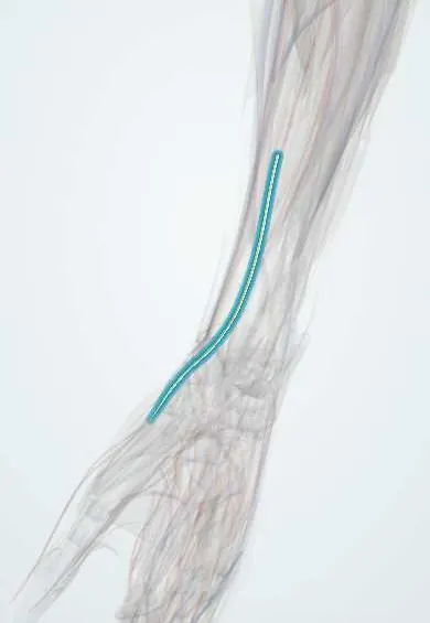
Palmar cutaneous branch !
The palmar cutaneous branch leaves the median nerve above the wrist; it gives feeling to the base of the palm and is spared in carpal tunnel syndrome.
Official name: Palmar cutaneous branch of the median nerve
Opens in the Durgical app, no sign-up needed.
What it is
A small skin branch that leaves the median nerve above the wrist and passes over the carpal tunnel.
What it does
Feeling in the skin of the base of the palm; it is spared in carpal tunnel syndrome.
Key facts
- Roots
- Median nerve, leaves above the wrist
- Moves
- None
- Feeling from
- Skin of the central palm and thenar eminence; spared in carpal tunnel syndrome
Relations
- Course
- Runs down between the tendons of flexor carpi radialis and palmaris longus, pierces the forearm fascia just above the wrist and passes over the flexor retinaculum to fan out over the thenar eminence and central palm
- Lateral
- The flexor carpi radialis tendon
- Medial
- The palmaris longus tendon
- Deep
- The flexor retinaculum, which separates it from the median nerve in the carpal tunnel
- Superficial
- Skin and the palmar aponeurosis
How to find it
Not palpable. Its path is marked by the interval between the flexor carpi radialis and palmaris longus tendons, both made visible by flexing the wrist against resistance; it lies just ulnar to the flexor carpi radialis tendon at the wrist crease.
Examination
- Pinprick over the thenar eminence and central palm: loss together with median finger numbness places a median lesion above the wrist; isolated loss after surgery suggests injury to this branch
- Tinel's sign over a tender scar at the palm base reproducing tingling into the thenar skin suggests a neuroma of this branch
On imaging
Not seen on radiographs. High-frequency ultrasound can follow it between the flexor carpi radialis and palmaris longus tendons, lying on top of the flexor retinaculum. After a palm-base scar, a neuroma appears as a small dark (hypoechoic) swelling continuous with the branch. The same scan checks the median nerve in the carpal tunnel. MRI is rarely needed.
Clinical notes
- Carpal tunnel release incisions are kept in line with the radial border of the ring finger, ulnar to the thenar crease, to stay clear of this branch
- In the volar approach to the distal radius, staying radial to the flexor carpi radialis tendon protects it; dissection ulnar to that tendon puts it at risk
- A painful neuroma can follow wrist ganglion excision or lacerations at the base of the palm
Anatomical variants
- Its height of origin on the median nerve varies and it may be doubled; when it is absent, the lateral antebrachial cutaneous or superficial radial nerve takes over the thenar skin
- Occasionally it runs through the flexor retinaculum or the palmaris longus tendon sheath instead of above them, where it is more exposed in surgery
Test yourself
Think of the answer, then tap the question to see it.
Where does the palmar cutaneous branch of the median nerve cross the wrist?
Superficial to the flexor retinaculum, not through the carpal tunnel, after leaving the median nerve a few centimetres above the wrist crease.
Why is thenar skin sensation spared in carpal tunnel syndrome?
The thenar skin and palm base are supplied by the palmar cutaneous branch of the median nerve, which passes over the flexor retinaculum.
How is the palmar cutaneous branch of the median nerve protected in carpal tunnel release?
The incision stays in line with the radial border of the ring finger, ulnar to the thenar crease.
Educational information, not medical advice. For any symptom, see a health professional.
Its names
- Official Latin (TA2)
- ramus palmaris nervi mediani TA2 6462
- Official Spanish (DeCS)
- Ramo palmar del nervio mediano
- English (TA2)
- Palmar cutaneous branch of the median nerve
- Also called
- palmar cutaneous branch of median nerve
Related
Sources: Terminologia Anatomica (TA2); Anatomy, Shoulder and Upper Limb, Median Nerve; Anatomy, Shoulder and Upper Limb, Wrist Flexor Retinaculum.
Spotted a mistake?We are always improving for the community. Tell us what is wrong and our team will review it.Report a mistake
Representative model, for educational purposes only.