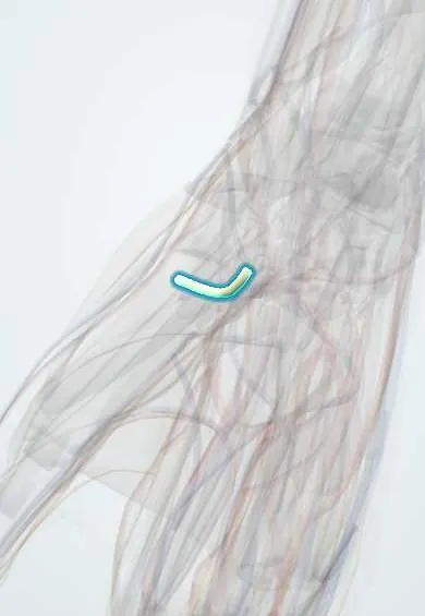
Recurrent motor branch !
The recurrent motor branch of the median nerve curls into the thumb pad; it moves the thumb-pad muscles, and surgeons protect it in carpal tunnel release.
Official name: Recurrent (thenar) motor branch of the median nerve
Opens in the Durgical app, no sign-up needed.
What it is
A short branch that curls back from the median nerve into the thumb pad, just past the carpal tunnel.
What it does
Powers the thumb-pad muscles; surgeons protect it during carpal tunnel release.
Key facts
- Roots
- Median nerve (C8–T1), just distal to the flexor retinaculum
- Moves
- APB, opponens pollicis, superficial head of FPB
- Feeling from
- None
Relations
- Course
- Leaves the median nerve at or just beyond the distal edge of the flexor retinaculum, turns upward and laterally (hence 'recurrent') and enters the thenar muscles near the border between abductor pollicis brevis and flexor pollicis brevis
- Superficial
- The palmar aponeurosis and the thenar fascia
- Medial
- The common palmar digital branches of the median nerve, which continue into the palm
How to find it
Not palpable. Its entry into the thenar muscles is marked by Kaplan's cardinal line, drawn from the apex of the first web space (thumb fully abducted) toward the hook of the hamate: the branch lies about where this line meets a line down the radial border of the middle finger.
Examination
- Pen test: with the hand flat and palm up, the thumb is raised straight up to touch a pen held above the palm; weakness indicates loss of abductor pollicis brevis from this branch or the median nerve
- Opposition: touching the thumb tip to the little finger pad with the thumbnail parallel to the palm; failure or weakness indicates opponens pollicis loss
- Inspection for thenar wasting, best seen from the side with both hands together
On imaging
Too small for radiographs. High-frequency ultrasound may show it curving back from the distal edge of the flexor retinaculum into the thenar muscles. In practice the median nerve is assessed instead: on ultrasound it looks swollen just before the carpal tunnel in compression. Long-standing loss of this branch leaves thenar atrophy, seen on MRI as fatty replacement of the muscles.
Clinical notes
- Divided by a carpal tunnel incision that strays radially, or by deep cuts over the thenar eminence; repair or a tendon transfer (opponensplasty) can restore opposition
- In carpal tunnel syndrome, nerve conduction records motor fibres of this branch over abductor pollicis brevis; a prolonged distal motor latency supports the diagnosis
Anatomical variants
- Its path relative to the flexor retinaculum varies: extraligamentous (most common), subligamentous, or transligamentous through the retinaculum, the type most at risk in surgery
- It may leave the ulnar side or the front of the median nerve and cross it, or be doubled
Memory aid
Thenar muscles it supplies, 'OAF' (the median 'LOAF' without the lumbricals): Opponens pollicis, Abductor pollicis brevis, Flexor pollicis brevis.
Related conditions
Test yourself
Think of the answer, then tap the question to see it.
Why is the recurrent motor branch of the median nerve called the «million-dollar nerve»?
Cutting it during carpal tunnel surgery loses thumb opposition.
Which muscles does the recurrent motor branch of the median nerve supply?
Abductor pollicis brevis, opponens pollicis and the superficial head of flexor pollicis brevis.
Which course of the recurrent motor branch relative to the flexor retinaculum is most at risk in surgery?
The transligamentous type, passing through the retinaculum; the extraligamentous course is the most common.
How can some thenar function survive a median nerve injury?
Through a Riche-Cannieu anastomosis between the recurrent motor branch and the deep branch of the ulnar nerve.
Educational information, not medical advice. For any symptom, see a health professional.
Its names
- Official Latin (TA2)
- ramus recurrens nervi mediani TA2 6464
- Official Spanish (DeCS)
- Ramo recurrente del nervio mediano
- English (TA2)
- Recurrent (thenar) motor branch of the median nerve
- Also called
- thenar motor branch
Related
Sources: Terminologia Anatomica (TA2); Anatomy, Shoulder and Upper Limb, Median Nerve; Anatomy, Shoulder and Upper Limb, Wrist Flexor Retinaculum.
Spotted a mistake?We are always improving for the community. Tell us what is wrong and our team will review it.Report a mistake
Representative model, for educational purposes only.