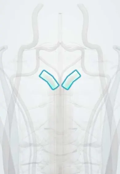
Alar ligaments !
The alar ligaments are two short, strong cords from the tip of the dens to the skull; they limit turning and tilting of the head.
Opens in the Durgical app, no sign-up needed.
What it is
Two short, strong cords from the tip of the dens to the sides of the skull opening.
What it does
Limit head rotation and side bending at the top of the neck; can be injured in whiplash.
Key facts
- Attaches
- Sides of the dens tip to the inner sides of the occipital condyles
- Function
- Check ligaments of the craniovertebral junction; limit head rotation and side bending; can tear in whiplash
- Tight in
- Head rotation (contralateral ligament) and lateral flexion
Relations
- Medial
- The apical ligament of the dens, a thin midline cord between the two alar ligaments, running from the dens tip to the front rim of the foramen magnum
- Behind
- Superior band of the cruciform ligament, then the tectorial membrane, the dura and the junction of the medulla and the spinal cord
- Below
- The dens of the axis (C2) and the median atlantoaxial joint, with the transverse ligament of the atlas wrapped behind the dens
Examination
- Alar ligament stress test (manual therapy): the examiner holds the C2 spinous process and side-bends the head; with an intact ligament C2 moves almost at once, while free side-bending before C2 moves suggests laxity. Not done in acute trauma until fracture and instability are ruled out on imaging
On imaging
They cannot be felt. Thin-slice MRI (coronal and axial, proton density or T2) shows two dark bands running up and out from the dens tip to the occipital condyles; a gap or high signal suggests a tear. CT shows avulsion fragments from the dens tip or the condyle.
Common problems
- Weakened in rheumatoid arthritis or Down syndrome, or torn by trauma: C1-C2 instability that can press on the cord.
How it is diagnosed
Flexion-extension X-rays (the gap between the atlas and the dens), CT and MRI.
Treatment
- Surgical C1-C2 fusion if the joint is unstable.
Clinical notes
- A dens-tip (type I) avulsion is usually stable by itself, but it can be a sign of a wider craniocervical (occipitocervical) injury
- In whiplash, alar ligament damage has been proposed as a cause of chronic upper neck pain; the MRI findings are debated
- In atlantoaxial instability from rheumatoid arthritis, they loosen together with the transverse ligament of the atlas
Memory aid
Alar means wing: two short wings spread out from the tip of the dens to the skull.
Test yourself
Think of the answer, then tap the question to see it.
What are the attachments of the alar ligaments?
From the sides of the tip of the dens (C2) to the inner sides of the occipital condyles: two short, strong cords.
Which head movements do the alar ligaments limit?
Rotation and side bending at the top of the neck: the ligament of one side checks rotation of the head to the opposite side.
Which fracture of the dens is an avulsion at the alar ligament attachment?
Type I odontoid fracture (Anderson–D'Alonzo), at the dens tip; usually stable by itself, but it can signal a wider craniocervical injury.
Can the alar ligaments hold the atlas in place if the transverse ligament of the atlas has failed?
No. They are only a secondary restraint to forward slip of C1 and cannot hold it on their own once the transverse ligament has failed.
Educational information, not medical advice. For any symptom, see a health professional.
Its names
- Official Latin (TA2)
- ligamenta alaria TA2 1695
- Official Spanish (DeCS)
- Ligamentos alares
- English (TA2)
- Alar ligaments
- Also called
- Mauchart's ligaments
Related
Sources: Terminologia Anatomica (TA2); Anatomy, Head and Neck: Atlantoaxial Joint; Anatomy, Head and Neck: Foramen Magnum.
Spotted a mistake?We are always improving for the community. Tell us what is wrong and our team will review it.Report a mistake
Representative model, for educational purposes only.