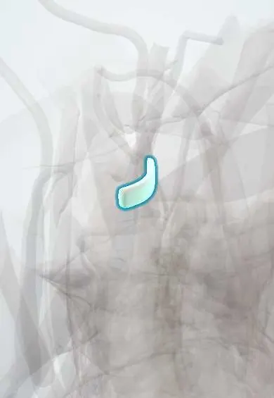
Transverse ligament of the atlas !
The transverse ligament of the atlas crosses the inside of the C1 ring and holds the dens of C2 forward, away from the spinal cord.
Official name: Transverse ligament of atlas
Opens in the Durgical app, no sign-up needed.
What it is
A strong band across the inside of the atlas ring, holding the dens of C2 against the front arch.
What it does
Keeps the dens from moving backward into the spinal cord, while letting the atlas turn around it.
Key facts
- Attaches
- Tubercles on inner sides of atlas lateral masses, passing behind the dens; with superior and inferior bands forms the cruciform ligament
- Function
- Holds dens against anterior arch of atlas; stops the dens moving back into the cord; laxity or rupture (e.g. rheumatoid arthritis) causes atlantoaxial instability
- Tight in
- Head and neck flexion (resists forward slide of atlas on axis)
Relations
- In front
- The dens, with a small synovial joint between the dens and the ligament: the back part of the median atlantoaxial joint
- Behind
- Tectorial membrane, dura and the spinal cord at the C1 level
- Above
- Superior band of the cruciform ligament to the front rim of the foramen magnum, behind the apical ligament and the alar ligaments
- Below
- Inferior band of the cruciform ligament to the back of the body of the axis (C2)
Examination
- Sharp–Purser test: seated, with the neck slightly flexed, the examiner fixes the C2 spinous process and gently pushes the forehead back; a clunk or easing of symptoms as the atlas slides back over the dens suggests atlantoaxial instability. Done with great caution, never in acute trauma; imaging is the safer first step
On imaging
Lateral flexion-extension radiographs measure the ADI and the posterior atlantodental interval (space left for the cord); in rheumatoid arthritis a posterior interval under about 14 mm is linked to cord risk. MRI shows the ligament directly as a dark band behind the dens. CT shows avulsion fragments from the C1 lateral mass.
Common problems
- Weakened in rheumatoid arthritis or Down syndrome, or torn by trauma: C1-C2 instability that can press on the cord.
How it is diagnosed
Flexion-extension X-rays (the gap between the atlas and the dens), CT and MRI.
Treatment
- Surgical C1-C2 fusion if the joint is unstable.
Clinical notes
- Jefferson fracture (burst of the C1 ring from an axial load): stability depends mainly on whether this ligament is intact
- In odontoid fractures the ligament usually stays intact, but the broken dens moves with the atlas, so the canal can still narrow
- Rheumatoid arthritis: pannus from the joint behind the dens erodes and loosens it; anterior atlantoaxial subluxation is the most common pattern
- Down syndrome: ligament laxity; screening flexion-extension radiographs have been used before some sports, though their value is debated
Memory aid
Cruciform means cross: the transverse ligament is the horizontal arm, the superior and inferior bands are the vertical arm.
Related conditions
Test yourself
Think of the answer, then tap the question to see it.
Where does the transverse ligament of the atlas attach, and which ligament does it help form?
To tubercles on the inner sides of the atlas lateral masses, passing behind the dens; with its superior and inferior bands it forms the cruciform ligament.
Which atlantodental interval (ADI) in an adult suggests injury to the transverse ligament of the atlas?
More than about 3 mm; a much wider gap suggests the alar ligaments have failed too.
What is Steel's rule of thirds at the level of the atlas?
The canal at C1 holds one-third dens, one-third spinal cord and one-third free space.
In a Jefferson fracture, what decides stability, and which X-ray rule suggests the transverse ligament is torn?
Whether the transverse ligament is intact; on the open-mouth view, C1 lateral masses overhanging C2 by about 7 mm or more in total (rule of Spence) suggest rupture.
How does rheumatoid arthritis affect the transverse ligament of the atlas?
Pannus from the joint behind the dens erodes and loosens it; anterior atlantoaxial subluxation is the most common pattern.
Educational information, not medical advice. For any symptom, see a health professional.
Its names
- Official Latin (TA2)
- ligamentum transversum atlantis; ligamentum cruciforme TA2 1703
- Official Spanish (DeCS)
- Ligamento transverso del atlas
- English (TA2)
- Transverse ligament of atlas
- Also called
- transverse atlantal ligament · cruciate ligament of atlas
Related
Sources: Terminologia Anatomica (TA2); Anatomy, Head and Neck: Atlantoaxial Joint; Anatomy, Head and Neck: Foramen Magnum.
Spotted a mistake?We are always improving for the community. Tell us what is wrong and our team will review it.Report a mistake
Representative model, for educational purposes only.