Cardiovascular system · Head and neck
Anterior jugular vein !
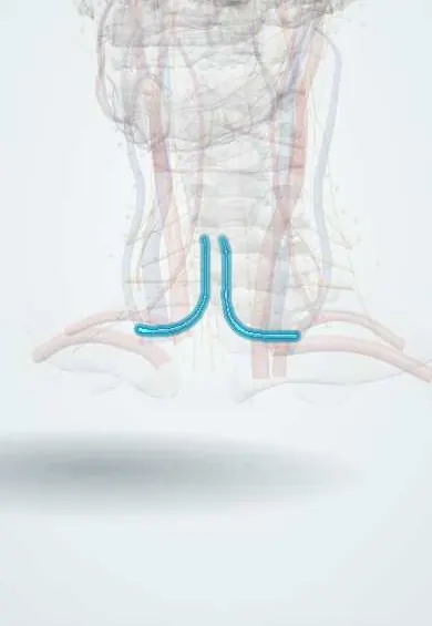
The anterior jugular vein is a small vein running down the front of the neck near the midline; it can bleed during a tracheostomy.
Opens in the Durgical app, no sign-up needed.
What it is
A small vein near the midline of the front of the neck.
What it does
Can bleed during tracheostomy.
Key facts
- Drains
- Front of the neck below the chin
- Into
- External jugular or subclavian vein (jugular venous arch links both)
Relations
- Course
- Begins below the chin near the hyoid bone from small submental veins, descends a little lateral to the midline over the strap muscles, then turns outward deep to the sternocleidomastoid just above the clavicle
- Superficial
- Skin, superficial fascia and, in its upper part, the platysma
- Deep
- Infrahyoid (strap) muscles, the sternohyoid and sternothyroid, with the larynx, thyroid gland and trachea beneath them
- Below
- Just above the sternum it runs in the suprasternal space between layers of the investing fascia, where the jugular venous arch crosses from side to side
How to find it
Often not visible. In a thin neck it may be seen a finger's width or two from the midline, filling when the person strains. In surgery it is met right after the skin and platysma, before the strap muscles are separated along the midline.
On imaging
Ultrasound before a percutaneous tracheostomy or cricothyroidotomy maps the anterior jugular veins and the jugular venous arch over the trachea so the puncture avoids them; colour Doppler confirms flow. On contrast CT they lie just under the platysma beside the midline, in front of the strap muscles, and can enlarge as collateral routes when deeper veins are blocked.
Clinical notes
- Tracheostomy: dissection stays in the midline between the two veins; the jugular venous arch low in the neck may need to be tied off
- Can be torn when skin flaps are raised in thyroid surgery or in penetrating injuries of the front of the neck
- It can enlarge as a collateral when the internal jugular or brachiocephalic vein is blocked (for example by a catheter-related clot), and a wide jugular venous arch makes low midline surgery bloodier
Anatomical variants
- The paired veins may be replaced by a single median vein of the neck
- Its size tends to vary inversely with that of the external jugular vein: when one is large, the other is often small
Test yourself
Think of the answer, then tap the question to see it.
What links the two anterior jugular veins low in the neck?
The jugular venous arch, above the sternum in the suprasternal space.
Why is the anterior jugular vein a concern in tracheostomy?
It and the jugular venous arch lie in the path of a midline tracheostomy and are a common source of bleeding.
Where does the anterior jugular vein end?
It turns laterally deep to the sternocleidomastoid to end in the external jugular or subclavian vein.
Educational information, not medical advice. For any symptom, see a health professional.
Its names
- Official Latin (TA2)
- vena jugularis anterior TA2 4959
- Official Spanish (DeCS)
- Vena yugular anterior DeCS D007601
- English (TA2)
- Anterior jugular vein
- Also called
- vena jugular superficialis ventralis
Sources: Terminologia Anatomica (TA2); DeCS; Anatomy, Head and Neck, External Jugular Veins; Anatomy, Head and Neck: Anterior Cervical Region.
Spotted a mistake?We are always improving for the community. Tell us what is wrong and our team will review it.Report a mistake
Representative model, for educational purposes only.