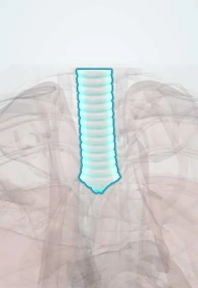
Trachea !
The trachea, or windpipe, is the tube of cartilage rings that carries air from the throat into the chest, where it divides into the two bronchi.
Opens in the Durgical app, no sign-up needed.
What it is
The windpipe: a tube about 10-12 cm long held open by C-shaped rings of cartilage, running from the larynx to its split (the carina) behind the sternum.
What it does
Carries air to and from the lungs; its lining moves mucus up with tiny hairs.
Key facts
- Where
- Midline from C6 (lower cricoid) to bifurcation at the sternal angle (T4–T5, carina); in front of the esophagus
- Blood supply
- Inferior thyroid arteries (upper part); bronchial arteries (lower part)
- Nerve
- Vagus via recurrent laryngeal nerves (sensory, parasympathetic); sympathetic trunk
- Function
- Air passage to the lungs; 16–20 C-shaped cartilage rings, open behind and closed by trachealis muscle
Relations
- In front
- Neck: thyroid isthmus over the 2nd to 4th rings, infrahyoid muscles, inferior thyroid veins; chest: manubrium, thymus, left brachiocephalic vein, aortic arch, brachiocephalic trunk and left common carotid artery
- Behind
- Esophagus, with the recurrent laryngeal nerves in the tracheoesophageal groove on each side; then the vertebral bodies
- Lateral
- Neck: thyroid lobes and carotid sheaths; chest, right: right vagus nerve, arch of the azygos vein, right pleura and lung; left: aortic arch, left common carotid and left subclavian arteries, left recurrent laryngeal nerve
- Below
- Divides at the carina into the main bronchi; the subcarinal lymph nodes fill the angle between them, and the left atrium lies just below
How to find it
It is felt in the midline of the neck from the cricoid cartilage down to the suprasternal (jugular) notch; a fingertip in the notch checks that the gap on each side of the trachea is equal. The carina projects at the sternal angle, where the 2nd costal cartilage meets the sternum, and moves lower in deep inspiration.
Examination
- Tracheal tug (Oliver sign): the examiner lifts the cricoid cartilage gently with the patient's chin raised; a downward pull with each heartbeat suggests an aortic arch aneurysm pressing on the left main bronchus
- Tracheal descent in inspiration (Campbell sign; some texts also call it tracheal tug): the larynx moves down toward the sternum with each breath in severe airflow obstruction such as COPD
On imaging
On a frontal chest X-ray the air column should sit midline over the spinous processes. The tip of an endotracheal tube is placed a few centimetres above the carina (commonly taught 5 ± 2 cm with the neck neutral). A widened carinal angle suggests an enlarged left atrium or subcarinal nodes. CT shows the lumen in COPD, where it can narrow side to side (saber-sheath trachea).
Common problems
- Narrowing after a long time with a breathing tube.
- Inhaled objects, especially in children.
How it is diagnosed
CT, bronchoscopy.
Treatment
- Bronchoscopy to remove objects; stents or surgery for narrowings.
Clinical notes
- A tracheostomy placed too low risks the brachiocephalic trunk and left brachiocephalic vein, which cross in front of the lower trachea; erosion into the trunk causes massive bleeding
- Emergency cricothyroidotomy goes through the cricothyroid membrane, above the trachea, because it is quicker and less vascular than a tracheostomy
- A large goitre or mediastinal mass can compress or displace the trachea, seen on the X-ray and felt at the suprasternal notch
Anatomical variants
- Tracheal bronchus: a bronchus to the right upper lobe, or to one of its segments, that leaves the trachea above the carina; usually found by chance on CT or bronchoscopy
Development
Forms from the respiratory diverticulum of the foregut, which the tracheoesophageal septum separates from the esophagus. Incomplete separation gives esophageal atresia with tracheoesophageal fistula; the commonest form is a blind upper esophagus with a lower fistula into the trachea.
Related conditions
- Foreign-body airway obstruction (choking)
- Drowning
- Follicular, oncocytic (Hürthle cell) and anaplastic thyroid carcinoma
- Influenza (flu)
- COVID-19
- Healthcare-associated infections (CLABSI, CAUTI, VAP)
- Croup (laryngotracheobronchitis)
- Pertussis (whooping cough)
- Foreign body aspiration in children
- Esophageal atresia and tracheoesophageal fistula
- Hospital-acquired and ventilator-associated pneumonia
- Acute bronchitis
- Major trauma (polytrauma): ATLS primary survey
- Burns (thermal, chemical, electrical; inhalation injury)
- Hypothyroidism
- Laryngeal cancer
- Thyroid nodule
- Thyroid cancer (papillary)
Test yourself
Think of the answer, then tap the question to see it.
Where does the trachea begin and where does it divide?
It begins at C6 below the cricoid cartilage and divides at the carina at the level of the sternal angle (T4–T5).
Through which tracheal rings is a tracheostomy usually made, and which structure crosses them?
The 2nd to 4th rings, which lie behind the thyroid isthmus; the isthmus is divided or retracted.
Why is a low tracheostomy dangerous?
The brachiocephalic trunk and left brachiocephalic vein cross in front of the lower trachea; erosion into the trunk causes massive bleeding.
Which way does the trachea shift in tension pneumothorax and in lobar collapse?
Away from a tension pneumothorax or massive effusion, and toward a collapsed lobe or pneumonectomy; checked at the suprasternal notch.
What lies behind the trachea, and what runs in the groove between them?
The esophagus, behind the cartilage-free wall closed by the trachealis muscle; the recurrent laryngeal nerves run in the tracheoesophageal grooves.
Educational information, not medical advice. For any symptom, see a health professional.
Its names
- Official Latin (TA2)
- trachea; carina tracheae TA2 3213
- Official Spanish (DeCS)
- Tráquea DeCS D014132
- English (TA2)
- Trachea
Related
Sources: Terminologia Anatomica (TA2); DeCS; Anatomy, Head and Neck, Trachea; Anatomy, Thorax, Tracheobronchial Tree.
Spotted a mistake?We are always improving for the community. Tell us what is wrong and our team will review it.Report a mistake
Representative model, for educational purposes only.