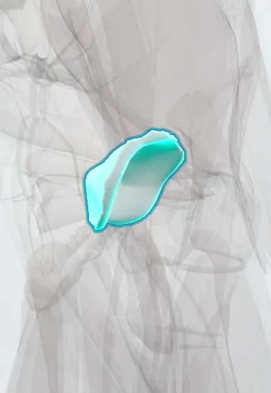
Acetabular cartilage !
The acetabular cartilage is the smooth, horseshoe-shaped lining of the hip socket, thickest at the roof where weight is borne.
Official name: Articular cartilage of acetabulum
Opens in the Durgical app, no sign-up needed.
What it is
Articular cartilage lining the hip socket. It is horseshoe-shaped, with a bare area in the middle filled by fat and the ligament of the femoral head.
What it does
Gives the ball a smooth surface to glide on and spreads the body's weight across the socket.
Key facts
- Where
- Horseshoe-shaped lunate surface of acetabulum; acetabular fossa and notch have none (fat pad, ligament of head)
- Function
- Hyaline articular cartilage; low-friction gliding; spreads load; avascular, nourished by synovial fluid
Relations
- Lateral
- Femoral head cartilage across the joint; the labrum continues the rim beyond the cartilage edge
- Medial
- Acetabular fossa, free of cartilage, with its fat pad and the ligament of the head; the quadrilateral plate beyond
- Below
- Acetabular notch, where the horseshoe opens, bridged by the transverse acetabular ligament
- Deep
- Subchondral bone of the roof, seen on X-ray as the dense sourcil
How to find it
Not palpable. At arthroscopy the central compartment is opened with traction and the cartilage is mapped by clock-face zones; the anterosuperior zone is damaged most often.
On imaging
AP pelvis: joint-space width at the roof, sourcil slope (Tönnis angle) and centre-edge angle for dysplasia. Judet views or CT for acetabular fractures; MR arthrography or cartilage mapping for delamination.
Common problems
- Hip osteoarthritis: gradual wear of the cartilage, causing groin or thigh pain, stiffness and a limp.
- Cartilage damage from hip impingement, dysplasia or injury.
- Avascular necrosis: loss of blood supply to the femoral head, which can make the bone and its cartilage collapse.
How it is diagnosed
Symptoms and exam (painful, limited inward turning of the hip); X-rays show joint-space narrowing and bone spurs; MRI shows early cartilage damage or avascular necrosis.
Treatment
- Physical therapy, weight management, a cane and activity changes; medicines or injections for pain.
- Hip arthroscopy for selected cartilage or labrum problems in younger hips without advanced arthritis.
- Total hip replacement for advanced arthritis: the worn ball and socket are replaced with artificial parts.
Clinical notes
- Periacetabular (Ganz) osteotomy reorients the socket in dysplasia before arthritis sets in
- A step in the roof after an acetabular fracture leads to post-traumatic arthritis
- Protrusio acetabuli: the femoral head bulges medial to the ilioischial line, seen in rheumatoid arthritis and some connective tissue disorders
Development
The ilium, ischium and pubis meet in the socket at a Y-shaped (triradiate) cartilage, which closes in the teens (texts differ on the age). Injury to this cartilage in a child can stop the socket from deepening and leave a shallow, dysplastic acetabulum.
Related conditions
Test yourself
Think of the answer, then tap the question to see it.
What shape is the acetabular cartilage, and which part has none?
Horseshoe-shaped (lunate surface); the acetabular fossa and notch have none, holding a fat pad and the ligament of the head.
Where is the acetabular cartilage thickest?
At the roof (superior dome), the main weight-bearing zone.
Why does acetabular dysplasia damage the rim cartilage early?
The shallow roof concentrates load on the rim, so rim cartilage and the labrum fail early.
Educational information, not medical advice. For any symptom, see a health professional.
Its names
- Official Latin (TA2)
- cartilago articularis TA2 382
- Official Spanish (DeCS)
- Cartílago articular del acetábulo DeCS D002358
- English (TA2)
- Articular cartilage of acetabulum
Related
Sources: Terminologia Anatomica (TA2); DeCS; Anatomy, Bony Pelvis and Lower Limb, Hip Joint; 8.3 The Pelvic Girdle and Pelvis (OpenStax (Rice University), 2022) · adapted, CC BY 4.0.
Spotted a mistake?We are always improving for the community. Tell us what is wrong and our team will review it.Report a mistake
Representative model, for educational purposes only.