Joints and ligaments · Lower limb
Acetabular labrum !
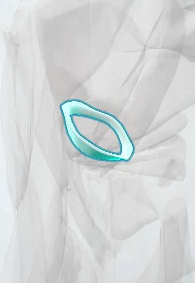
The acetabular labrum is a ring of cartilage on the rim of the hip socket; it deepens the socket and seals the joint.
Opens in the Durgical app, no sign-up needed.
What it is
A ring of tough cartilage attached to the rim of the hip socket (acetabulum).
What it does
Deepens the socket, helps hold the ball in place, and seals the joint so the joint fluid keeps the cartilage cushioned.
Key facts
- Where
- Fibrocartilage ring on the acetabular rim; bridges the acetabular notch as the transverse acetabular ligament
- Attaches
- Bony acetabular rim; transverse acetabular ligament
- Function
- Deepens socket; grips femoral head beyond its equator; seals joint fluid (suction seal); adds stability
Relations
- Lateral
- Joint capsule, attached just beyond it, leaving a narrow perilabral recess
- Medial
- Femoral head cartilage, which it grips beyond the equator; acetabular cartilage at the chondrolabral junction
- In front
- The iliopsoas tendon crosses in front of the anterior capsule and labrum and can damage it (iliopsoas impingement)
- Above
- The reflected head of rectus femoris attaches to the ilium and capsule just above the superior labrum
- Below
- Transverse acetabular ligament across the acetabular notch, under which vessels enter the fossa
How to find it
Not palpable. At hip arthroscopy it is seen in the central compartment between the femoral head and the acetabular rim. Tears are mapped like a clock face, with 12 o'clock superior and 3 o'clock anterior.
Examination
- FADIR test (flexion, adduction, internal rotation): reproduced groin pain suggests impingement or a labral tear; sensitive but not specific
- FABER (Patrick) test: groin pain points to the hip joint, posterior pain to the sacroiliac joint
- Log roll: rolling the straight leg in and out; pain or a click suggests an intra-articular source
On imaging
MR arthrography is the reference test: contrast entering the labrum or its base marks a tear; normal sublabral sulci can mimic one. X-rays look for the cause: cam (alpha angle on a lateral view), pincer (crossover sign, deep socket) and dysplasia (lateral centre-edge angle).
Common problems
- Labral tears from hip impingement (FAI), hip dysplasia, twisting sports or wear with age.
- Symptoms: groin pain, clicking, catching or locking, and stiffness.
- A torn labrum can speed up wear of the joint cartilage.
How it is diagnosed
Physical exam (impingement tests that bend the hip and turn it inward); X-rays to look at bone shape; MR arthrogram (MRI with contrast in the joint). A numbing injection into the joint can confirm that the pain comes from inside the hip.
Treatment
- Without surgery: activity changes, physical therapy, anti-inflammatory medicine, and sometimes an injection.
- Hip arthroscopy to repair or trim the labrum, and to treat the bone shape that caused the tear.
- Labral reconstruction with a graft from the patient or a donor when the labrum cannot be repaired.
Clinical notes
- C sign: a person with intra-articular hip pain often cups the side of the hip with thumb and fingers to show where it hurts
- In hip dysplasia the rim is overloaded, so the labrum enlarges and tears
- At arthroscopy the labrum is repaired or trimmed, usually together with reshaping of the bone that caused the tear
Anatomical variants
- A sublabral sulcus, a normal cleft between the labrum and the acetabular cartilage, can look like a tear on MR arthrography; studies differ on its usual site and how often it occurs
Development
Like the capsule, it arises from mesenchyme around the interzone of the embryonic hip. In developmental dysplasia the rim can be everted or turned into the socket (the limbus), where it stops the femoral head from seating concentrically.
Related conditions
Test yourself
Think of the answer, then tap the question to see it.
Where do most acetabular labral tears occur, and why?
Anterosuperiorly: the zone loaded in flexion and internal rotation, where cam and pincer impingement act.
Which test reproduces groin pain with flexion, adduction and internal rotation in impingement or a labral tear?
The FADIR test; it is sensitive but not specific.
What is the reference imaging test for an acetabular labral tear?
MR arthrography: contrast entering the labrum or its base marks a tear (normal sublabral sulci can mimic one).
Why does the articular side of the acetabular labrum heal poorly?
Most of the blood supply reaches the capsular (outer) side; the articular side is poorly vascularised.
Educational information, not medical advice. For any symptom, see a health professional.
Its names
- Official Latin (TA2)
- labrum acetabuli TA2 1880
- Official Spanish (DeCS)
- Labrum acetabular
- English (TA2)
- Acetabular labrum
- Also called
- glenoid labrum of hip · labrum acetabulare · labrum glenoidale · labium articulare
Related
Sources: Terminologia Anatomica (TA2); Anatomy, Bony Pelvis and Lower Limb, Hip Joint; 9.7 Development of Joints (OpenStax (Rice University), 2022) · adapted, CC BY 4.0.
Spotted a mistake?We are always improving for the community. Tell us what is wrong and our team will review it.Report a mistake
Representative model, for educational purposes only.