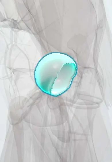
Femoral head cartilage !
The femoral head cartilage is the smooth layer covering the ball of the hip joint; it thins and wears away in hip arthritis.
Official name: Articular cartilage of femoral head
Opens in the Durgical app, no sign-up needed.
What it is
Articular cartilage: a smooth, slippery layer covering the ball (head) of the femur.
What it does
Lets the ball glide in the socket with very little friction and spreads the body's weight across the joint.
Key facts
- Where
- Femoral head, except the fovea (where the ligament of the head attaches)
- Function
- Hyaline articular cartilage; low-friction gliding; spreads load; avascular, nourished by synovial fluid
Relations
- Above
- Acetabular cartilage of the lunate surface, with the labrum around the rim
- Medial
- Fovea, free of cartilage, with the ligament of the head and its artery; the fat pad of the acetabular fossa
- Below
- Head–neck junction where the cartilage ends; a cam bump sits at its anterosuperior part
- Deep
- Subchondral bone, fed by retinacular branches of the medial circumflex femoral artery climbing along the neck
How to find it
Not palpable. The head lies deep to the femoral artery just below the midpoint of the inguinal ligament; its cartilage is seen at hip arthroscopy and in joint replacement.
On imaging
AP pelvis: superolateral narrowing, osteophytes, subchondral cysts and sclerosis in osteoarthritis; crescent sign then flattening in avascular necrosis (Ficat stages). MRI shows early avascular necrosis with a double-line sign and cartilage delamination.
Common problems
- Hip osteoarthritis: gradual wear of the cartilage, causing groin or thigh pain, stiffness and a limp.
- Cartilage damage from hip impingement, dysplasia or injury.
- Avascular necrosis: loss of blood supply to the femoral head, which can make the bone and its cartilage collapse.
How it is diagnosed
Symptoms and exam (painful, limited inward turning of the hip); X-rays show joint-space narrowing and bone spurs; MRI shows early cartilage damage or avascular necrosis.
Treatment
- Physical therapy, weight management, a cane and activity changes; medicines or injections for pain.
- Hip arthroscopy for selected cartilage or labrum problems in younger hips without advanced arthritis.
- Total hip replacement for advanced arthritis: the worn ball and socket are replaced with artificial parts.
Clinical notes
- Chondrolysis, a rapid loss of joint cartilage, is a recognised complication of slipped capital femoral epiphysis
- Risk factors for avascular necrosis include corticosteroids, alcohol, sickle cell disease, decompression illness and hip fracture or dislocation
Development
The femoral head is all cartilage at birth, so ultrasound, not X-ray, is used to screen young infants for hip dysplasia. Its ossification centre appears in the first year (often around 4–6 months; some texts later), and late appearance is a sign of dysplasia; its growth plate closes in mid adolescence.
Related conditions
- Hip Osteoarthritis
- Slipped capital femoral epiphysis
- Legg-Calvé-Perthes disease
- Avascular necrosis of the femoral head
- Transient synovitis of the hip
Test yourself
Think of the answer, then tap the question to see it.
Which part of the femoral head has no cartilage?
The fovea, where the ligament of the head attaches.
How does joint-space narrowing differ between hip osteoarthritis and inflammatory arthritis?
Osteoarthritis narrows it typically superolaterally; inflammatory arthritis narrows it evenly (axial or concentric).
What is the crescent sign in avascular necrosis of the femoral head?
A thin lucent line under the cartilage of the dome: a subchondral fracture just before collapse.
Educational information, not medical advice. For any symptom, see a health professional.
Its names
- Official Latin (TA2)
- cartilago articularis TA2 382
- Official Spanish (DeCS)
- Cartílago articular de la cabeza del fémur DeCS D002358
- English (TA2)
- Articular cartilage of femoral head
Related
Sources: Terminologia Anatomica (TA2); DeCS; Anatomy, Bony Pelvis and Lower Limb, Hip Joint; 8.5 Development of the Appendicular Skeleton (OpenStax (Rice University), 2022) · adapted, CC BY 4.0.
Spotted a mistake?We are always improving for the community. Tell us what is wrong and our team will review it.Report a mistake
Representative model, for educational purposes only.