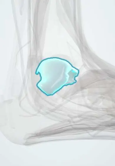
Talar cartilage !
The talar cartilage is the smooth layer covering the joint surfaces of the ankle bone (talus), so the ankle glides.
Official name: Articular cartilage of talus
Opens in the Durgical app, no sign-up needed.
What it is
Articular cartilage: a smooth, slippery layer covering the faces of the talus where it meets the tibia, the fibula and the heel bone.
What it does
Lets the ankle glide with very little friction and spreads the body's weight across the bone.
Key facts
- Where
- Trochlea (dome) and malleolar facets of talus; also subtalar and talonavicular surfaces (about 60% of talus covered)
- Function
- Hyaline articular cartilage; low-friction gliding; spreads load; avascular, nourished by synovial fluid
Relations
- Above
- Tibial plafond cartilage
- Medial
- Articular facet of the medial malleolus
- Lateral
- Articular facet of the lateral malleolus of the fibula
- Below
- Posterior, middle and anterior calcaneal facets of the subtalar joint
- In front
- Head facet for the navicular
- Deep
- Subchondral bone with a limited blood supply
How to find it
The front of the dome is partly felt in front of the ankle with the foot fully plantarflexed. Anterolateral lesions lie near this area; posteromedial ones are reached in surgery through a posterior approach or an osteotomy of the medial malleolus.
On imaging
The mortise view may show a lucency or a flake on the dome, but many lesions are missed on X-ray. MRI shows bone oedema and whether the fragment is stable; CT shows its size and bony depth for planning.
Common problems
- Osteochondral lesions: a damaged patch of cartilage and the bone under it, often after an ankle sprain or fracture.
- Ankle arthritis, often years after an injury.
- Symptoms: deep ankle pain, swelling, catching or stiffness.
How it is diagnosed
Symptoms and exam; X-rays; MRI or CT show the size and depth of a lesion.
Treatment
- Rest, a boot and physical therapy, especially for small or stable lesions.
- Surgery for focal lesions: cleaning the area and making small holes in the bone to start healing (microfracture), or cartilage repair and grafting with bone and cartilage from the patient or a donor (osteochondral graft).
- Advanced arthritis: ankle fusion or ankle replacement.
Clinical notes
- Small stable lesions are often treated by bone marrow stimulation; larger ones by osteochondral grafts
- Subtalar cartilage damage after a calcaneal fracture leads to subtalar arthritis
Development
Like other joint surfaces it is hyaline cartilage kept from the embryonic cartilage model of the talus; in adolescents, osteochondritis dissecans of the talar dome is one cause of defects in this layer.
Related conditions
Test yourself
Think of the answer, then tap the question to see it.
How do lateral and medial talar dome osteochondral lesions classically differ?
Lateral: shallow, wafer-like, usually post-traumatic; medial: deeper, cup-shaped, less often linked to injury.
What is the most common cause of ankle osteoarthritis?
Previous trauma; ankle cartilage resists degeneration better than the knee's, so ankle osteoarthritis is mostly post-traumatic.
Which imaging best shows whether a talar dome osteochondral fragment is stable?
MRI, which shows bone oedema and fragment stability; many lesions are missed on X-ray.
Educational information, not medical advice. For any symptom, see a health professional.
Its names
- Official Latin (TA2)
- cartilago articularis TA2 382
- Official Spanish (DeCS)
- Cartílago articular del astrágalo (tróclea) DeCS D002358
- English (TA2)
- Articular cartilage of talus
Related
Sources: Terminologia Anatomica (TA2); DeCS; Anatomy, Bony Pelvis and Lower Limb: Ankle Joint; 9.7 Development of Joints (OpenStax (Rice University), 2022) · adapted, CC BY 4.0.
Spotted a mistake?We are always improving for the community. Tell us what is wrong and our team will review it.Report a mistake
Representative model, for educational purposes only.