Ankle fracture !
An ankle fracture is a break of one or both bony bumps of the ankle (the malleoli), often with torn ligaments that can make the joint unstable.
Updated 2026-10-29
Structures involved
Tap one to see what it is and open it in 3D.
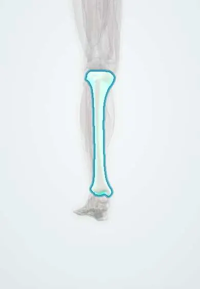
- Tibia
- Fibula
- Talus
- Syndesmosis ligaments
- Deltoid ligament
- ATFL
- Tibial plafond cartilage
- Talar cartilage
- Superficial fibular nerve
- Dorsalis pedis artery
In the clinical exam
What it is
A break of one or more of the malleoli (the lower ends of the tibia and fibula), often with ligament injury that can make the ankle joint unstable.
Who gets it
One of the most common lower-limb fractures. Low-energy twisting injuries predominate; peaks are seen in young active men and in older women.
Causes
- Twisting (rotational) injury of the foot on the leg
- Falls, sport and stepping off a kerb
- High-energy trauma (road traffic, falls from height)
Risk factors
- Osteoporosis and older age
- Obesity
- Contact and pivoting sports
- Diabetes with neuropathy (worse healing and complications)
- Smoking (delays bone and wound healing)
Pathophysiology
The ankle mortise is a ring of bone and ligaments around the talus. A rotational force breaks this ring at one or more points: lateral malleolus, medial malleolus or deltoid ligament, posterior malleolus and syndesmosis. If two parts of the ring fail, the talus can shift and the joint becomes unstable.
Classification
- Weber A: fibular fracture below the syndesmosis, usually stable
- Weber B: at the level of the syndesmosis, stability varies
- Weber C: above the syndesmosis, syndesmosis injured, usually unstable
- By number of malleoli: unimalleolar, bimalleolar, trimalleolar
- Lauge-Hansen classification by mechanism (foot position and force direction)
Symptoms and signs
- Pain and swelling around the ankle after injury
- Inability to bear weight
- Bruising
- Tenderness over the malleoli
- Visible deformity when dislocated
Diagnosis
- Clinical
- The Ottawa ankle rules guide the need for X-ray: bone tenderness at the back edge or tip of either malleolus, or inability to bear weight for four steps. Check the proximal fibula, foot pulses and nerves.
- Tests
- Usually none for the fracture; pre-operative tests when surgery is planned.
- Imaging
- Ankle X-rays (AP, lateral and mortise views) show the fracture and talar shift. Stress views judge stability; CT for complex or posterior malleolus fractures.
Differential diagnosis
- Lateral ankle sprain
- High ankle (syndesmosis) sprain
- Fifth metatarsal base fracture
- Talar or calcaneal fracture
- Achilles tendon rupture
Goal of treatment
Restore a congruent, stable ankle mortise so the talus sits centred, allowing the joint to heal with good function and lower arthritis risk.
Treatment
- Acute
- Urgent reduction and splinting of a dislocated ankle to protect skin, vessels and nerves.
- Conservative
- Stable fractures (often Weber A and many isolated Weber B) are treated in a boot or cast with early weight bearing as tolerated.
- Surgical
- Unstable, displaced or bimalleolar/trimalleolar fractures and syndesmosis injuries are usually treated with open reduction and internal fixation.
- Rehabilitation
- Physiotherapy for range of motion, strength, balance and return to walking.
Complications
- Post-traumatic ankle arthritis
- Malunion or non-union
- Wound problems and infection, especially in diabetes or smokers
- Deep vein thrombosis
- Stiffness and chronic swelling
- Complex regional pain syndrome
Prognosis
Most stable fractures heal well. Outcomes depend on restoring the mortise; residual talar shift or syndesmosis malreduction raises the risk of arthritis.
Prevention
- Fall prevention and bone health in older adults
- Balance and proprioception training in sport
Test yourself
Think of the answer, then tap the question to see it.
Which findings make an ankle X-ray necessary under the Ottawa ankle rules?
Bone tenderness at the back edge or tip of the lateral or medial malleolus, or inability to bear weight for four steps both at injury and in the clinic.
In the Weber classification, where is the fibular fracture in a type C injury?
Above the syndesmosis; the syndesmosis is injured and the ankle is usually unstable.
What does a trimalleolar fracture involve?
The lateral malleolus, the medial malleolus and the posterior malleolus (the back of the distal tibia).
Why is the proximal fibula examined in an ankle injury?
A Maisonneuve injury (high fibular fracture with syndesmosis and medial injury) can be missed if only the ankle is imaged.
Related conditions
Educational information, not medical advice. For any symptom, see a health professional.
Sources: Ankle Fracture (StatPearls (NCBI Bookshelf), 2025).
Spotted a mistake?We are always improving for the community. Tell us what is wrong and our team will review it.Report a mistake
Representative model, for educational purposes only.