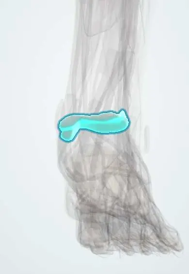
Tibial plafond cartilage !
The tibial plafond cartilage is the smooth layer on the lower end of the shin bone, forming the roof of the ankle joint.
Official name: Articular cartilage of tibial plafond
Opens in the Durgical app, no sign-up needed.
What it is
Articular cartilage covering the lower end of the tibia (the plafond), the roof of the ankle joint.
What it does
Lets the ankle glide with very little friction and spreads the body's weight across the bone.
Key facts
- Where
- Inferior articular surface of distal tibia (plafond), continuous with medial malleolus facet; roof of ankle mortise
- Function
- Hyaline articular cartilage; low-friction gliding; spreads load; avascular, nourished by synovial fluid
Relations
- Below
- Talar dome cartilage
- Medial
- Continuous with the articular facet of the medial malleolus
- Lateral
- Fibular notch and the syndesmosis with the lateral malleolus
- In front
- Anterior tibial lip and capsule; the anterior tibial artery and deep fibular nerve cross in front of the ankle between the tendons
- Behind
- Posterior malleolus, attachment of the posterior inferior tibiofibular ligament; flexor hallucis longus behind
- Deep
- Subchondral bone of the distal tibia
How to find it
Not palpable. The joint line lies a little above the tip of the medial malleolus; it is palpable just medial and lateral to the extensor tendons; in anterior ankle arthroscopy the plafond is the roof seen above the talar dome.
On imaging
Mortise view for joint-space symmetry; CT for pilon fracture planning (Rüedi–Allgöwer types by comminution and joint displacement); MRI for cartilage lesions.
Common problems
- Damage from fractures of the lower tibia (pilon fractures) or from ankle sprains.
- Ankle arthritis, often years after an injury.
How it is diagnosed
Symptoms and exam; X-rays; MRI or CT show the size and depth of a lesion.
Treatment
- Rest, a boot and physical therapy, especially for small or stable lesions.
- Surgery for focal lesions: cleaning the area and making small holes in the bone to start healing (microfracture), or cartilage repair and grafting with bone and cartilage from the patient or a donor (osteochondral graft).
- Advanced arthritis: ankle fusion or ankle replacement.
Clinical notes
- A large posterior malleolus fragment (Volkmann fragment) involves the back of the plafond and affects stability and congruity
- Tillaux fracture in adolescents: the anterior inferior tibiofibular ligament avulses the anterolateral distal tibial epiphysis while the lateral part of the growth plate is still open
- Severe pilon fractures are often treated in stages, with a temporary frame until the soft tissues settle
Development
The distal tibial growth plate closes from its central-medial part outwards over about 18 months in adolescence, which explains Tillaux and triplane fractures.
Related conditions
Test yourself
Think of the answer, then tap the question to see it.
In a pilon fracture, what largely sets the later risk of ankle arthritis?
Cartilage damage at impact, as the talus is driven into the tibial plafond.
On the mortise view, what does a medial clear space wider than the space under the plafond mean?
Talar shift; normally the joint space is even all round.
What is a Tillaux fracture?
In adolescents, the anterior inferior tibiofibular ligament avulses the anterolateral distal tibial epiphysis while the lateral growth plate is still open.
Educational information, not medical advice. For any symptom, see a health professional.
Its names
- Official Latin (TA2)
- cartilago articularis TA2 382
- Official Spanish (DeCS)
- Cartílago articular de la superficie articular inferior de la tibia DeCS D002358
- English (TA2)
- Articular cartilage of tibial plafond
Related
Sources: Terminologia Anatomica (TA2); DeCS; Anatomy, Bony Pelvis and Lower Limb: Ankle Joint; Anatomy of the Human Body (20th ed.): Surface Anatomy of the Lower Extremity (Lea & Febiger (Gray's Anatomy, 1918), 1918) · adapted, Public domain; Anatomy of the Human Body (20th ed.): Surface Markings of the Lower Extremity (Lea & Febiger (Gray's Anatomy, 1918), 1918) · adapted, Public domain.
Spotted a mistake?We are always improving for the community. Tell us what is wrong and our team will review it.Report a mistake
Representative model, for educational purposes only.