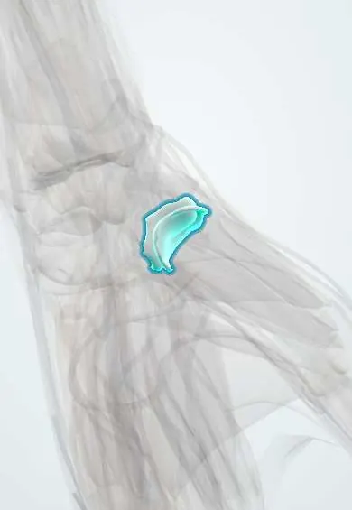
Thumb base cartilage !
The thumb base cartilage covers the saddle joint between the trapezium and the thumb's metacarpal; it often wears out with age (thumb base arthritis).
Official name: Cartilage of the thumb carpometacarpal (CMC) joint
Opens in the Durgical app, no sign-up needed.
What it is
The saddle-shaped cartilage between the trapezium and the thumb metacarpal.
What it does
Lets the thumb swing across the palm; it often wears out with age (thumb base arthritis).
Key facts
- Where
- Saddle surfaces of the trapezium and the base of the 1st metacarpal
- Function
- Saddle joint allows opposition; common site of osteoarthritis
Relations
- Above
- Trapezium, whose saddle surface faces distally and radially
- Below
- Base of the 1st metacarpal
- Lateral
- APL tendon inserting on the metacarpal base; the radial artery crosses the floor of the anatomical snuffbox nearby on its way to the first web space
- In front
- Thenar muscles (abductor pollicis brevis, opponens pollicis); the FCR tendon runs in its groove on the trapezium just medially
- Behind
- Dorsoradial ligament and branches of the superficial radial nerve
How to find it
Sliding a finger up the back of the first metacarpal toward the wrist, the step where it meets the trapezium, at the far end of the anatomical snuffbox, is the joint; it glides under the finger as the thumb moves. In advanced arthritis the base sits prominent and the thumb base looks squared.
On imaging
A true AP (Robert) view, taken with the hand fully pronated, shows the joint without overlap; the Eaton–Littler stages (I–IV) grade narrowing, osteophytes and involvement of the scaphotrapeziotrapezoid (STT) joint.
Common problems
- Thumb base arthritis (rhizarthrosis), very common in women over 50.
- Pain at the base of the thumb when pinching, opening jars or turning keys; later a bump and a stiff, narrowed web.
How it is diagnosed
Pain with the grind test at the thumb base. X-rays show the joint narrowing and spurs.
Treatment
- Splint, hand therapy and joint-protection techniques; anti-inflammatory medicines.
- Steroid injection for flare-ups.
- Surgery (removing the trapezium with a tendon sling, joint fusion or replacement) when other treatment fails.
Clinical notes
- Rolando fracture: a comminuted (Y- or T-shaped) intra-articular fracture of the first metacarpal base, with a worse outlook than a Bennett fracture
- Surgery for advanced basal joint arthritis includes trapeziectomy, with or without ligament reconstruction and tendon interposition, or joint replacement or fusion
- De Quervain tenosynovitis (positive Finkelstein test) and STT arthritis are the main look-alikes for pain at the thumb base
Development
Unlike the other metacarpals, whose secondary centre is at the head, the first metacarpal has it at the base, so its growth plate lies right next to this joint, as in a phalanx.
Test yourself
Think of the answer, then tap the question to see it.
Why is the thumb carpometacarpal joint a prime site for arthritis?
Its two reciprocal saddles have little bony constraint, so stability depends on ligaments.
Which ligament holds the small fragment in a Bennett fracture?
The palmar beak (anterior oblique) ligament, while the abductor pollicis longus pulls the shaft away.
Which conditions are the main look-alikes for pain at the thumb base?
De Quervain tenosynovitis (positive Finkelstein test) and STT arthritis.
Educational information, not medical advice. For any symptom, see a health professional.
Its names
- Official Latin (TA2)
- cartilago articularis articulationis carpometacarpeae pollicis TA2 382
- Official Spanish (DeCS)
- Cartílago articular de la articulación carpometacarpiana del pulgar
- English (TA2)
- Cartilage of the thumb carpometacarpal (CMC) joint
- Also called
- thumb basal joint cartilage
Related
Sources: Terminologia Anatomica (TA2); Anatomy, Shoulder and Upper Limb, Hand Bones.
Spotted a mistake?We are always improving for the community. Tell us what is wrong and our team will review it.Report a mistake
Representative model, for educational purposes only.